


Serviços Personalizados
Artigo
 Inglês (pdf)
 Artigo em XML
 Referências do artigo
Indicadores

Links relacionados
-
 Citado por Google
 -
 Similares em Google
Compartilhar

 Permalink
South African Dental Journal
versão On-line ISSN 0375-1562
versão impressa ISSN 0011-8516
S. Afr. dent. j. vol.72 no.3 Johannesburg Abr. 2017
CLINICAL REVIEW
Lodox® digital imaging - A tool for dental identification in single and mass fatality situations
H BernitzI; J VersterII
IBChD, MSc, PhD. Senior Stomatologist, Department of Oral Pathology and Oral Biology, Faculty of Health Sciences, University of Pretoria, South Africa
IIMBChB, MMed (Forens Path), FC For Path (SA). Senior Lecturer / Specialist Forensic Pathologist, Department of Forensic Medicine, Faculty of Health Sciences, University of Pretoria, South Africa
ABSTRACT
Lodox®Statscan™ is a relatively recent addition to the field of post- mortem radiology and imaging in forensic dentistry and forensic medicine. A number of cases were encountered where victims who could not undergo visual identification were positively identified by comparing their post mortem Lodox® images with their ante-mortem dental records. A brief description is provided of how Lodox®Statscan™ images can be of benefit in cases where visual identification is not feasible or must be validated by another method of post-mortem identification. The digital images also provide a permanent record of the dentition of the deceased for future reference. The emphasis of this article is on the Lodox®Statscan™ as a screening tool for cases which are suitable for dental identification, especially in mini-mass fatality and mass fatality situations, which is a potent new possibility in the field of dental identification. The use of these full body low dosage digital radiographic images for the purposes of dental identification has not been reported in the literature previously.
Keywords: Forensic Dentistry; Lodox®Statscan™; full body scan; low dosage radiation; dental identification.
INTRODUCTION
Lodox®Statscan™ (LS) was developed by the South African mining industry in order to perform low dosage regular screening of employees to detect illegal diamond smuggling.1-4 During 1999 it was realized that this low dose digital scanner is an effective triage tool in emergency medicine departments in hospitals, as full body images could be obtained in a matter of seconds, with minimal manipulation of the trauma victim.5-9 The LS is an equally valuable application in forensic medicine, and amongst the advantages discussed in the literature are the detection of peripheral skeletal injuries and foreign objects, such as projectiles, especially in multiple gunshot injury cases.3-4,7,9 Lodox has been applied as a screening device for maxillofacial injuries,1 but exploring the application of Lodox digital radiographs in forensic odontology, especially for the purpose of dental identification, has to date been neglected in the dental literature.
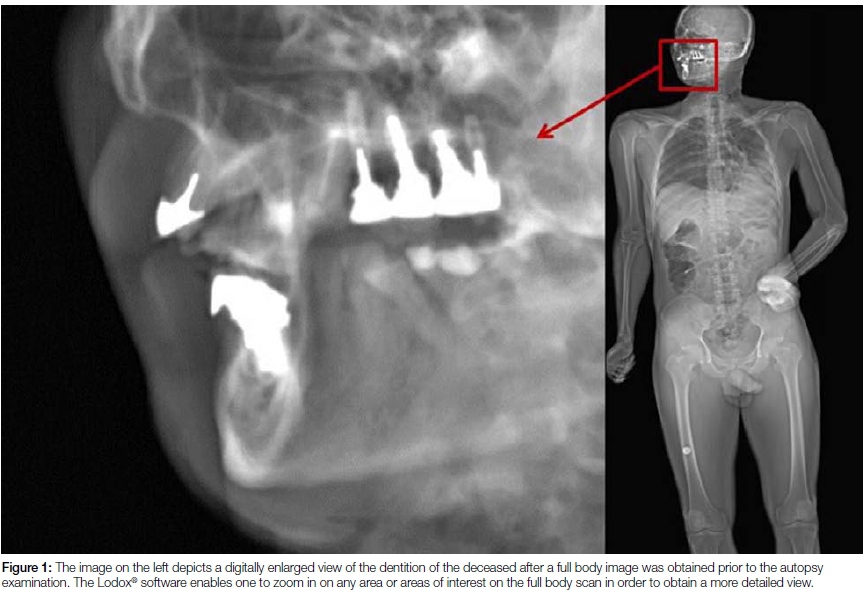
Many forensic investigative facilities do not have continuous access to a forensic odontologist for expert consultation nor do they have advanced dental imaging modalities available. LS could provide a fast primary radiographic survey of bodies admitted for medico-legal investigation, especially in cases where the identification of the deceased is unknown.5 Cases with recognisable dental restorations can be selectively separated for dental identification. In South Africa, a country with financial constraints, limited staff and restricted availability of post mortem radiographic imaging modalities, LS provides an alternative to obtaining full body digital recordings of each deceased individual admitted to our medico-legal laboratories. The system also enables radiographic screening for unique dental features. Any aspect of the digital image, including the dentition of the deceased, can be enlarged and digitally enhanced to render a more detailed picture.6
LS is a useful imaging tool in medico-legal laboratories with high case loads, as it can obtain a full body lateral and anterior-posterior scan in an average of only 5-6.5 minutes.6,7It has the ability to scan bodies in quick succession,6 emits low radiation, has minimal scatter of radiation and thus can be placed directly in the autopsy room/operational area, without putting staff at additional risk. Although it is difficult to compare the effective dose of radiation of a full body anteroposterior (AP) scan with the effective dose of conventional radiographs, it has been shown that the effective dose emitted by a Lodox® full body AP scan performed on a medium sized adult is approximately 14% of the effective dose of an abdominal AP image taken by a conventional X-ray machine.10 Similar findings were apparent in a paediatric study, where it was found that the effective doses of LS AP abdomen and AP pelvis scans comprised only 10% of the effective dose of similar images taken by conventional radiography.11 In fact, ten Lodox® full body scans per year would still expose the individual receiving those scans to less radiation than the prescribed maximum accidental dose to the general public of 1 millisievert (mSv) per year, as per the 1990 recommendations of the International Commission on Radiological Protection.8, 11 Another major advantage is that images can be viewed almost immediately after the scan is completed.6


It has been suggested that the LS could enable health care workers in the clinical setting to utilise radiographs in the mass disaster, "mini-mass disaster", or high volume trauma scenarios, as previously this would have been a time consuming and labour intensive exercise, with numerous logistical constraints.2 The use of LS for identification purposes in mass fatalities and specifically for dental identification has previously not been reported in the literature.
Two cases are used to illustrate the identification process using LS.
CASE 1
The presence of multiple concordant dental features with no inexplicable discrepancies when comparing the maxillary and mandibular dental arches on the LS with the ante-mortem dental records serves to confirm the identity.
The points of dental concordance included:
• 11. Porcelain veneer crown (position of the crown was checked in the anterior-posterior angulated image)
• 48 F am O
• 47 F am MO
• 46 F am O
• 45 F am DO
• All lower anterior teeth sound
• 18 F am O
• 28 F am O
(F = filling, am= amalgam, O = occlusal, M = mesial, D = distal)
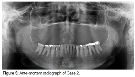
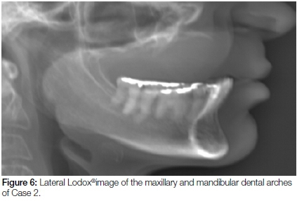
CASE 2
Comparison of the maxillary and mandibular dental arches of this case on the LS and the ante-mortem dental records also resulted in a positive dental identification.
The points of dental concordance included:
• Edentulous maxilla
• Root anatomy of the 47 similar
• Root position and anatomy of 46 similar
• 46 F am MOD similar
• 48 missing
• 37 F am MO (characteristic shape of amalgam similar)
• 36 F am MOD
(F = filling, am= amalgam, O = occlusal, M = mesial, D = distal)
In each case the forensic dentist was satisfied with the concordant features present for examination. Additional images may be taken if required, and can include skull rotations if necessary. In Case 1, it was deemed necessary to check that the crown present on the lateral radiograph was indeed the 21 and not any other anterior crown.
DISCUSSION
Lodox® scanners have successfully been used in our forensic mortuary to identify single victims and victims of multiple fatalities where decomposition, mutilation and/or carbonization have been present. Bodies are put through the LS to determine if any dental restorations or recognisable dental features are present. In each case where ante-mortem dental records were available, the LS images were compared with the ante-mortem records received from the dental surgeons and comparisons made. The quality of the images observed in the LS is sufficient to ensure a positive correlation, and thus an identification of the victim. As illustrated in the LS images, dental restorations, implants, composite restorations and tooth relationships are clearly visible. As a standard protocol all teeth and dental restorations are charted on Interpol F2 forms and odontograms constructed for the identification process. From our experience, the complete identification process should not take longer than 10 minutes per body. The two cases are included to illustrate the clarity of the LS and comparison of ante-mortem and post mortem records. It must be emphasised that when the head is being scanned laterally it must be tilted to avoid superimposition of the left and right sides (oblique lateral).
The use of the LS in mass disaster situations offers the following advantages.
1. The LS can facilitate the quick and effective identification of burned, decomposed and mutilated bodies in mass disasters. Dental identification that is quick, cheap and accurate is the method of choice when fingerprints and DNA are not available and visual identification is unreliable due to decomposition, mutilation or burning.
2. Dental identification can be carried out without any form of facial tissue damage. In cases of carbonisation or rigor mortis the dentition must be mechanically exposed to visualise the teeth. Conventional radiography requires the digital sensor to be placed in the palate or lingual to the mandibular teeth, necessitating access to an open oral cavity.
3. Obtaining radiographic images of the victim's dentition without having to gain surgical access to the oral cavity has benefits for certain religious groups.
4. Ability to carry out a dental examination before the start of the formal autopsy can facilitate the identification of key individuals e.g. the pilot/co-pilot.
5. In cases of severe carbonisation the tooth structures may become extremely fragile, and are often damaged or displaced during the exposure of the dentition. By using the LS it is possible to observe the dentition without any manipulation of the dental structures.
6. The detail observed in both the maxillary and mandib-ular dental arches is sufficient to perform age estimation analyses on children and young adults burned in mass disaster situations. In a recent aircraft accident, for example, three children aged 14, 9 and 6 were tentatively identified by radiological evaluation of their tooth development. This contributed greatly to their final identification.
7. A full body digital radiographic survey of each case can easily be saved as a permanent imaging record of the body. This can be used for comparison should dental records or other maxillofacial or skeletal identifying features captured on ante-mortem records become available at a later stage. These might include sinus patterns and orthopaedic prostheses.
8. Permanent full body digital images can be easily manipulated and enlarged and transferred to other institutions for consultation, especially in areas where no forensic odontologist is available.
The fact that the Lodox® scanner is not portable restricts the examinations of bodies to the main centres where the services are available. In practise it is often difficult to position bodies in rigor mortis, and more than one scan is required to ensure that the head is well aligned and that the extremities do not obscure either the facial features or the abdominal and pelvic cavities. Such procedures can prolong the time required to obtain the desired images. Although the Lodox provides diagnostic information similar to that of conventional X-rays, it cannot replace high resolution modalities like CT scanners and MRI-scanners. Lodox images cannot be used to compile 3D reconstructed images.
CONCLUSION
The routine use of LS at a medico-legal laboratory in Pretoria, South Africa, has proven that this imaging modality can be successfully utilized for the purposes of dental identification of severely mutilated victims, including charred and decomposed bodies.
All cases admitted to the medico-legal laboratory undergo a full body LS screening and the images are thus readily available for dental identification by the forensic odontologist where applicable.
Not only could LS digital radiographs be an effective method of conducting forensic dental identification, but serve also as a useful screening tool to detect the presence of dental work in victims of mass disasters.
References
1. Deyle S, Evangelopoulos DS, Brehmer T, Zimmermann H, Ex- adaktylos AK. Full body radiography (LODOX-Statscan) as a screening device for maxillofacial injuries, where CT scanning is not immediately available. Injury 2011; 42: 112-3. [ Links ]
2. Whiley SP, Alves H, Grace S. Full-body X-ray imaging to facilitate triage: a potential aid in high-volume emergency departments. Emerg Med Int 2013; 2013: 43: 70-8. [ Links ]
3. Fu CY, Wang YC, Hsieh CH, Chen RJ. Lodox/Statscan pro vides benefits in evaluation of gunshot injuries. Am J Emerg Med 2011; 29: 823-7. [ Links ]
4. Knobel GJ, Flash G, Bowie GF. Lodox/Statscan proves to be invaluable in forensic medicine. SAMJ 2008; 96: 593. [ Links ]
5. Chen RJ, Fu CY, Wu SC, Wang YC, Chung PK, Huang HC, Huang JC, Lu CW. Diagnostic accuracy, biohazard safety, and cost effectiveness-the Lodox/Statscan provides a ben eficial alternative for the primary evaluation of patients with multiple injuries. J Trauma 2010; 69: 826-30. [ Links ]
6. Boffard KD, Goosen J, Plani F, Degiannis E, Potgieter H. The use of low dosage X-ray (Lodox/Statscan) in major trauma: comparison between low dose X-ray and conventional X-ray techniques. J Trauma 2006; 60: 1175-83. [ Links ]
7. Deyle S, Brehmer T, Evangelopoulos DS, Krause F, Benneker LM, Zimmermann H, Exadaktylos AK. Review of Lodox/ Statscan in the detection of peripheral skeletal fractures in multiple injury patients. Injury 2010; 41: 818-22. [ Links ]
8. Exadaktylos AK, Benneker LM, Jeger V, Martinolli L, Bonel HM, Eggli S, Potgieter H, Zimmermann H. Total-body digital X-ray in trauma: an experience report on the first operational full body scanner in Europe and its possible role in ATLS. Injury 2008; 39: 525-9. [ Links ]
9. Evangelopoulos DS, Deyle S, Zimmermann H, Exadaktylos AK. Full-body radiography (Lodox/Statscan) in trauma and emergency medicine: a report from the first European installation site. Trauma 2011; 13: 5-15. [ Links ]
10. Irving BJ, Maree GJ, Hering ER, Douglas TS. Radiation dose from a linear slit scanning X-ray machine with full-body imaging capabilities. RadiatProtDosimetry 2008; 130: 482-9. [ Links ]
11. Maree GJ, Irving BJ, Hering ER. Paediatric dose measurement in a full-body digital radiography unit. Pediatr Radiol 2007; 37: 990-7. [ Links ]
 Correspondence:
Herman Bernitz
Department of Oral Pathology and Oral Biology, School of Dentistry, University of Pretoria
P.O. Box 1266
Pretoria, South Africa, 0001
Tel: +27 13 2826419, Cell: +27 845125258
E-mail: bernitz@iafrica.com
1 Statscan, Lodox Systems Pty (Ltd), Sandton, South Africa. LODOX is a loose acronym for "low dose X rays," according to Wikipaedia.

