


Services on Demand
Article
 English (pdf)
 Article in xml format
 Article references
Indicators

Related links
-
 Cited by Google
 -
 Similars in Google
Share

 Permalink
South African Dental Journal
On-line version ISSN 0375-1562
Print version ISSN 0011-8516
S. Afr. dent. j. vol.72 n.2 Johannesburg Mar. 2017
FORENSIC ODONTOLOGY
Forensic dentistry case book 8: Taking identification to a higher level
H BernitzI; G SaaymanII
IBChD., Dip (Odont)., MSc., PhD. (Pret). Department of Oral Pathology and Oral Biology, Faculty of Health Sciences, University of Pretoria
IIMBChB., MMed(MedForens) (Pret)., FCForPath(SA). Head, Department of Forensic Medicine, Faculty of Health Sciences, University of Pretoria
Dental identification of decomposed, burned and mutilated remains usually involves comparing the ante-mortem with the post-mortem dental records of the suspected victim. This procedure is dependent on the availability of usable ante-mortem records obtained from the respective dentist or dental specialist.1
This suicide case involved a gunshot wound to the mouth. The individual had pointed the barrel towards his mouth, destroying the tooth crowns in the pre-maxilla area. Many of the posterior teeth which were unaffected by the shooting were however restored with amalgam fillings and root treatments were observed on three of the molars. The ante-mortem records received from the dentist contained hand written notes describing a three unit porcelain veneer bridge which he had placed from the 12 to the 21. He stated that there was a root treatment on the first lower left molar, but had no other dental records or radiographs of the patient. The explanation given was that he had only done the anterior bridge and had noticed the root canal during that treatment. The digital radiographs had been destroyed in a thunder storm and no backups had been made.
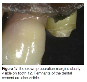
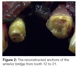
Although a root treatment was present on the 36 that was not sufficient for a positive identification to be made in this case without a radiograph of the tooth. Two days after the initial dental examination it was decided to revisit the body and see whether any clues could be found regarding the anterior bridge. The roots of the two anchor teeth were still intact, but no sign of the bridge was present on visual examination. The pre-maxilla was then re-examined under magnification. The magnified images of the two roots, after the mucosa had been retracted, clearly showed the margins of the bridge preparations. Figure 1 shows the prepared crown margin of tooth 12 as well as cement remnants on the prepared surface. The two pieces of the pre-maxilla were repositioned and showed that a bridge had been present before the shooting, see Figure 2. The presence of a root treatment on the 36, a missing 11, and crown margin preparations on the 12 and 21 were sufficient to positively identify the individual. All the above features are regarded as special dentistry and are thus uncommon in the general population. The positive dental identification expedited the return of the body to the next of kin. The alternative methods, namely DNA and fingerprint analysis, generally take extended periods to analyse which would further add to the psychological trauma of the already grieving family.
This case clearly shows the need for dental practitioners to keep good dental records. How was it possible for a patient who had extensive dental work, that radiographs were not taken, and that an odontogram or descriptions of the root canal treatments were not recorded? Also highlighted is the need to make backups of digital radiographs. It was only the force of circumstances that led us to perform microscopic analysis on the root remains of the teeth suspected of being the bridge anchors.
Reference
1. Van Niekerk PJ., Bernitz H. Retrospective investigation of dental records used in forensic identification cases. South African Dental Journal 2003:58(3); 102-4. [ Links ]
 Correspondence:
Herman Bernitz:
Department of Oral Pathology and Oral Biology,
Faculty of Health Sciences, University of Pretoria.
E-mail: bernitz@iafrica.com

