


Serviços Personalizados
Artigo
 Inglês (pdf)
 Artigo em XML
 Referências do artigo
Indicadores

Links relacionados
-
 Citado por Google
 -
 Similares em Google
Compartilhar

 Permalink
South African Dental Journal
versão On-line ISSN 0375-1562
versão impressa ISSN 0011-8516
S. Afr. dent. j. vol.71 no.9 Johannesburg Out. 2016
RADIOLOGY CASE
Maxilo-facial radiology case 145
CJ Nortjé
BChD, PhD, ABOMR, DSc. Faculty of Dentistry, University of the Western Cape. E-mail: cnortje@uwc.ac.za
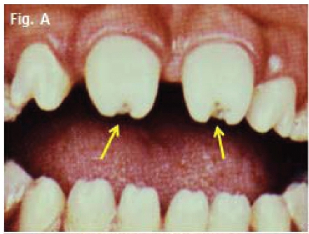
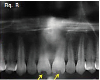
Below are clinical and radiographic images of patients who presented in the Department with the main complaint that they were not happy with the aesthetic appearance of some of their teeth which developed as they grew older. What is your diagnosis?
INTERPRETATION
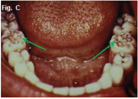
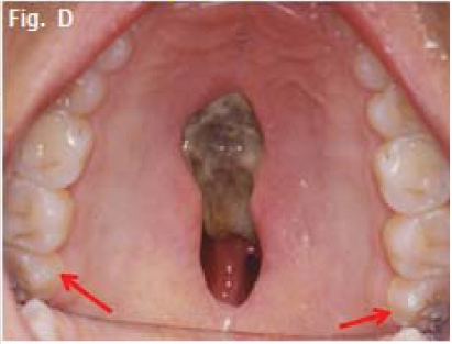
The images above present the classic features of congenital syphilis. Syphilis is caused by the infection with spirochete Treponema pallidum. The acquired form is usually further sub-classified into three distinctive stages: primary, secondary and tertiary. The bone may be affected in congenital syphilis and in both the secondary and tertiary stages of acquired syphilis. The jaws are rarely affected in syphilis. When they are, the palate is more frequently involved than is the mandible. The purpose of this communication is to discuss the effects of congenital syphilis on teeth. Figures A & B are examples of Hutchinson's teeth. Hutchinson's triad was first described by Jonathan Hutchinson, an English surgeon, in 1858 as being pathognomonic of congenital syphilis. The triad consisted of (1) diffuse interstitial keratitis (deep deposits in substance of the cornea, which becomes hazy throughout), (2) disease of the labyrinth (canals of the inner ear) and (3) Hutchinsonian teeth affecting the permanent maxillary incisors. The typical "Hutchinsonian incisor" is smaller than the normal incisor and the crown may converge from the cervix to the incisal edge. As a result, the tooth is narrower at the cutting edge than at the normal-sized gingival margin. This gives the tooth a barrel or screwdriver form. In addition the incisal edge is usually notched (Figs A &. B, yellow arrows). After Hutchinson's description, it was later recognized that changes might also occur in other permanent canines, and first molars. The "mulberry" molar (Fig.C) was first described by Fournier in 1884. Sometimes these molars are called Fournier molars or Moon's molars. Fig D is a case of congenital syphilis affecting the palate and demonstrating mulberry molars on the 18 and 28 (red arrows). According to Putkonen (1962), the so-called mulberry molar is smaller than the normal first molar tooth. The mulberry molar is covered on the sides with normal enamel but the occlusal surface is pinched together, dwarfed, rough, and hypoplastic, often pigmented (green arrows).
Reference
1. Hutchinson J: Report of effects of infantile syphilis in marring of teeth. Trans Pathol Soc (Lond) 9:449, 1858. [ Links ]
2. Fournier A: Syphilitic teeth. Dental Cosmos 26: 12, 81,141, 1884 [ Links ]
3. Farman AG, Nortje CJ & Wood R E: Oral and Maxillofacial Imaging, 1st Ed, Mosby. St. Louis, Missouri 1993 pp.204-205 [ Links ]
