


Serviços Personalizados
Artigo
 Inglês (pdf)
 Artigo em XML
 Referências do artigo
Indicadores

Links relacionados
-
 Citado por Google
 -
 Similares em Google
Compartilhar

 Permalink
South African Dental Journal
versão On-line ISSN 0375-1562
versão impressa ISSN 0011-8516
S. Afr. dent. j. vol.71 no.5 Johannesburg Jun. 2016
RADIOLOGY CASE
Maxillo-facial radiology case 141
CJ Nortjé
BChD, PhD, ABOMR, DSc. Faculty of Dentistry, University of the Western Cape. E-mail: cnortje@uwc.ac.za
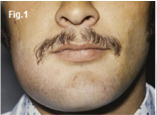
This thirty five year old male patient presented with a slow growing swelling in the right body of the mandible (Fig.1).The patient also experienced intermittent pain from time to time in the region of the swelling. What are the most important radiological features and what is your diagnosis?
INTERPRETATION
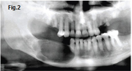
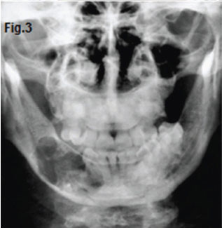
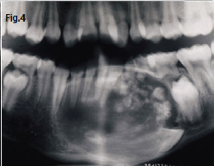
The cropped pantomograph and postero-anterior image (Figs.2 & 3) of the patient show a homogenous radiolucency in the right mandibular premolar and molar regions. Note the extensive calcification present in another case and the apparent dentigerous relationship to an impacted and displaced mandibular canine (Fig. 4). A histological diagnosis of a calcifying odontogenic cyst (COC) was made. The calcifying odontogenic cysts are developmental odontogenic lesions which are believed to arise from odontogenic epithelial remnants in the gingivae or in the mandible or maxilla. Over the years since its first description, it has become clear that the COC has a number of variants, including features of a benign odontogenic tumour. The COC was first described by Gorlin et al. (1962, 1964) who were impressed by the significant presence of so-called 'ghost cells' and its histological resemblance to the cutaneous calcifying epithelioma of Malherbe. The eponym of 'Gorlin cyst' is frequently used. Since its early description the lesion has been widely recognised, occurring both peripherally and centrally in the jaws, while its origins, pathogenesis and histopathological variations have evoked considerable discussion in the literature. Patients range widely in age (1 to 87 years) with a peak incidence for detection in the second decade of life. Some reports suggest that the cysts have a predilection for females, whereas other studies show no gender bias. More than 70% of COC are associated with the maxilla. The anterior segments of the jaw are most commonly affected. The calcifying odontogenic cyst may appear initially as a unilocular or multilolcular radiolucency with discrete, well demarcated margins. Irregular sized calcifications may be scattered in the radiolucency, producing variable degrees of opacity. This may produce a "salt and pepper" appearance. Denser opacities are likely to be present if the cyst is associated with a complex odontoma. The COC is treated by surgical enucleation unless it is associated with another odontogenic tumour in which case wider excision may be required.
Reference
1. Shear M. and Speight PM. (2007) Cysts of the Oral and Maxillofacial Regions, 4th edn.Blackwell, Munksgaard, pp100-108. [ Links ]
2. Farman AG, Nortje CJ & Wood R E: Oral and Maxillofacial Imaging, 1st Ed, Mosby. St. Louis, Missouri 1993 pp. 210-212 [ Links ]
