


Services on Demand
Article
 English (pdf)
 Article in xml format
 Article references
Indicators

Related links
-
 Cited by Google
 -
 Similars in Google
Share

 Permalink
South African Dental Journal
On-line version ISSN 0375-1562
Print version ISSN 0011-8516
S. Afr. dent. j. vol.71 n.5 Johannesburg Jun. 2016
CLINICAL REVIEW
Efficacy of Er,Cr:YSGG Laser in treatment of unusual presentation of Pyogenic Granuloma in a 9 year old girl
Karandeep S AroraI; Prabhpreet KaurII; Kasper D HundalIII; Rupinder D KaurIV; Shrawan K SharmaV
IAssistant Professor, Department of Oral Medicine & Radiology, Daswani Dental College & Research Centre, Kota, Rajasthan, India
IIAssistant Professor, Department of Oral & Maxillofacial Pathology, B.R.S. Dental College & General Hospital, Panchkula, Haryana, India
IIIPost Graduate Student, Department of Oral Medicine & Radiology Daswani Dental College & Research Centre, Kota, Rajasthan, India
IVPost Graduate Student, Department of Paedodontics & Preventive Dentistry,Daswani Dental College & Research Centre, Kota, Rajasthan, India
VPost Graduate Student, Department of Orthodontics & Dentofacial Othopaedics, Daswani Dental College & Research Centre, Kota, Rajasthan, India
ABSTRACT
The term pyogenic granuloma is a misnomer because the lesion does not contain pus and is not strictly speaking a granuloma. The growth is typically seen in young adults; however it may occur in any age, especially in individuals with poor oral hygiene. Some cases have also been reported in children. The latest treatment options include lasers of different type. Final diagnosis of the lesion is mainly by biopsy and histopathological examination. Here we report a case of pyogenic granuloma in a paediatric patient who was treated with Er,Cr:YSGG laser.
Keywords: Pyogenic granuloma, paediatric patient, inflammatory hyperplasia, lasers
INTRODUCTION
Pyogenic granuloma (PG) is one of the common inflammatory hyperplasis (IH) seen in the oral cavity.1 Hullihen's description in 1844 was most likely the first PG reported in English literature, but the term "pyogenic granuloma" or "granuloma pyogenicum" was introduced by Hartzell in 1904.2,3
The term "pyogenic granuloma" is a misnomer because the lesion does not contain pus and is not strictly speaking a granuloma. Approximately one-third of the lesions occur due to trauma and poor oral hygiene may also be one of the precipitating factors.4
Although it is a common disease in the skin, it is extremely rare in the gastrointestinal tract, except for the oral cavity where it is often found on keratinized tissue. There are two kinds of PG namely lobular capillary hemangioma (LCH type) and non-LCH type, which differ in their histological features.5-7
The growth is typically seen in young adults; however it may occur in any age, especially in individuals with poor oral hygiene. Females are far more susceptible than males because of the hormonal changes that occur in women during puberty, pregnancy, and menopause.8,9 Some cases have also been reported in children.10
Final diagnosis of the lesion is mainly by biopsy and histopathological examination. Conventional treatment of pyogenic granuloma consists of surgical excision along with elimination of irritating local factors. The latest treatment options include lasers of different type. Here we report an unusual case of pyogenic granuloma occurring on the gingiva of a paediatric patient,which was treated with Er,Cr:YSGG laser. This case is submitted after the prior approval of the Institutional Review Board.
CASE REPORT
A nine year old female child patient presented to the Department of Oral Medicine & Radiology with the chief complaint of a growth in the lower left posterior region of the mouth, first noticed by the parents of the patient about one month previously. The growth was initially smaller, had been gradual in onset and had progressively increased in size over the month. The patient and her parents were unaware of just when the lesion had first appeared. The gradual increase in size of the growth has caused discomfort while eating and there has also been occasional bleeding during chewing of food.
The past medical history was not contributory. Extra oral examination revealed no relevant findings.
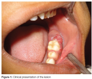
Intra oral examination revealed a solitary sessile growth situated behind tooth 36, involving the marginal gingiva and measuring 1.2 x 1 x 0.5 cms (Figure 1). The surface was smooth and erythematous with a groove in the centre, which was ulcerated, suggestive of indentations by the maxillary counterpart.
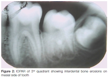
An intraoral periapical radiograph was taken of the 36, 37 region, which revealed slight interdental bone erosions on the mesial side of 37 (Figure 2).
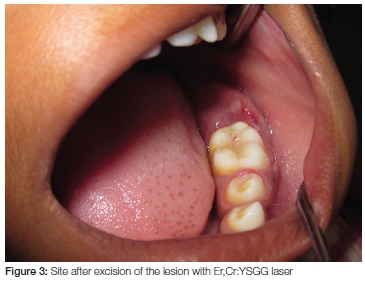
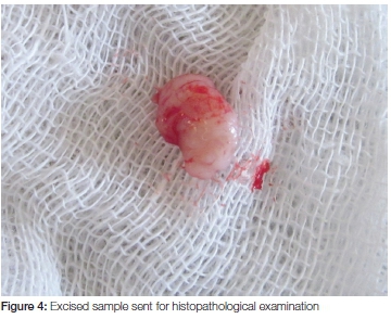
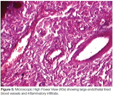
Blood investigations of the patient were performed, with results which were within normal range. An excisional biopsy was done under local anesthesia with a Er,Cr:YSGG laser with a wavelength of 2780nm. After the lesion was excised (Figure 3) the sample (Figure 4) was sent for histopathological examination (Figure 5).
DISCUSSION
The incidence of the pyogenic granuloma has been described as between 26.8% to 32% of all reactive lesions.11
According to Shafer et al., oral pyogenic granulomas arise as a result of infection by either staphylococci or streptococci, but also as a result of some minor trauma to the tissues that provides a pathway for invasion of nonspecific types of microorganisms. These authors explain the mechanism by suggesting that the tissue response invokes the well-known biologic principle that any irritant applied to living tissue may act either as a stimulus or as a destructive agent or both. If many cells are present in a small volume of tissue and there is a relative reduction of blood flow through the area, as in inflammation, the concentration of the stimulating substance will be high and growth will be stimulated. As differentiation and maturation are attained, the cells become widely separated and the concentration of the substance falls and little growth occurs. In the type of inflammation that results in the formation of oral pyogenic granuloma, destruction of fixed tissue cells is slight but the stimulus to proliferation of vascular endothelium persists and exerts its influence over a long period of time.12
Abdulai et al. in their retrospective study among 108 cases of oral pyogenic granuloma presenting in patients aged between 9 months to 71 years, concluded the peak ages of occurrence are 11 - 20 years with the commonest site being the gingivae (58.33%) ,and a higher prevalence in the upper jaw (42.59%). Other sites include the lips (18.52%), buccal mucosa (10.19%) and tongue (8.26%).10
Clinically pyogenic granuloma appears as a localized solitary lump having a sessile or pedunculated base. It is a well circumscribed benign soft tissue tumour of inflammatory rather than neoplastic nature arising from the connective tissue of the skin or mucous membrane.13 The surface can be smooth or lobulated, having a deep red or purplish colour. It is a vascularized lesion with a tendency to bleed profusely owing to micro trauma.4 In general there are no relevant radiographic findings in pyogenic granuloma. However, Angelopoulos in his review observed that localized alveolar bone resorption can be seen in rare instances of large and long standing gingival tumours.13
Histopathologically the major bulk of the lesion is formed by a non lobulated mass of angiomatous tissue. Usually, lobulated lesions are composed of solid endothelial proliferation or a proliferation of capillary sized blood vessels. Collagen in the connective tissue is sparse. The natural history of the lesion follows three distinct phases. In the cellular phase, the lobules are compact and cellular with little lumen formation. In the capillary phase the lobules become highly vascular with abundant intra-luminal red blood cells. In the involutionary phase there is a tendency for intra and perilobular fibrosis with increased venular differentiation. Increased vascularity may be noticed and some observers have reported that pyogenic granuloma is partly or completely covered by parakeratotic or non keratinized stratified squamous epithelium.4,14,15
Differential diagnosis of pyogenic granuloma includes parulis, peripheral giant cell granuloma, peripheral ossifying fibroma, leiomyoma, hemangioendothelioma, hemangiopericytoma, bacillary angiomatosis, Kaposi's sarcoma, metastatic tumour, pregnancy tumour and post extraction granuloma.6
For pyogenic granuloma, surgical excision is the treatment of choice. Another conventional surgical modality for treatment is cryosurgery in the form of either liquid nitrogen spray or cryoprobe. Nd:YAG and CO2 and flashlamp pulsed dye lasers have also been used for the treatment of oral pyogenic granuloma.16 Meffert et al. used the flash pulsed dye laser on a mass of granulation tissue that did not respond to the unusual treatment.17 Lasers have been shown to be a successful option for the excision of pyogenic granuloma with advantages of minimal pain and invasiveness and the lack of any need for suturing or packing. Dermal pyogenic granuloma has been treated with electrodessication and sclerotherapy.18 White et al have also suggested that laser excision is well accepted by patients with no adverse affects.
In the present case also Er,Cr:YSGG laser is a very precise ablation instrument that offers certain advantages. It is strongly absorbed by water and causes minimal damage to the adjacent tissues, especially the underlying muscle layers. In the present case, due to minimal trauma to the adjacent tissues, postoperative healing was favourable, with very little scar formation. Post operative bleeding in the case was minimal and no sutures were placed after the excision.
CONCLUSION
The present case was reported as it is unusual in the sense that it is occurring in a paediatric patient over the mucosa of an erupting tooth, and that treatment was with the use of lasers. This is an unusual combination, rarely reported in the literature.
ACRONYMS
IH: inflammatory hyperplasia
PG: pyogenic granuloma
LCH type: lobular capillary haemangioma and non-LCH
IOPAR: intra-oral periapical radiograph
References
1. Gondivkar SM, GadbailA, Chole R. Oral pregnancy tumour. Contemp Clin Dent 2010;1(3):190-2. [ Links ]
2. Hullihen SP. Case of aneurism by anastomosis of the superior maxillae. Am J Dent Sc 1844;4:160-2. [ Links ]
3. Hartzell MB. Granuloma pyogenicum. J Cutan Dis Syph 1904;22:520-5. [ Links ]
4. Bhaskar SN, Jacoway JR. Pyogenic granuloma: clinical features, incidence, histology, and result of treatment: report of 242 cases. J Oral Surg 1966;24(5):391-8. [ Links ]
5. Yao T, Nagai E, Utsunomiya T, Tsuneyoshi M. An intestinal counterpart of pyogenic granuloma of the skin. A newly proposed entity. Am J Surg Pathol 1995;19:1054-60. [ Links ]
6. Fowler EB, Cuenin MF, Thompson SH, Kudryk VL, Billman MA. Pyogenic granuloma associated with guided tissue regeneration: a case report. J Periodontol 1996;67:1011-5. [ Links ]
7. Epivatianos A, Antoniades D, Zaraboukas T, Zairi E, Poulopoulos A, Kiziridou A, Iordanidis S. Pyogenic granuloma of the oral cavity: comparative study of its clinicopathological and immunohistochemical features. Pathol Int 2005;55:391-7. [ Links ]
8. Neville BW, Damm DD, Allen CM, Bouquot JE. Oral & Maxillofacial Saunders; 2002. p. 437-5. [ Links ]
9. Regezi JA, Sciubba JJ, Jordan RCK. Oral Pathology: Clinical Pathologic Considerations. 4th ed, Philadelphia: WB Saunders; 2003. p. 115-6. [ Links ]
10. Abdulai AE, Nuamah IK, Baddoo H, Gyasi RK. Oral pyogenic granuloma in Ghanaians: a review of cases. International Journal of Medicine and Biomedical Research 2013;2(3):173-8. [ Links ]
11. Kfir Y, Buchner A, Hansen LS. Reactive lesions of the gingiva: A clinicopathologic study of 471 cases. J Periodontol 1980;51:655-61. [ Links ]
12. Shafer, Hine, Levy . Shafer's Textbook of Oral Pathology. 5th ed. Amsterdam: Elsevier Health Sciences; 2006. pp. 459-61. [ Links ]
13. Angelopolous AP. Pyogenic granuloma of the oral cavity: Statistical analysis of its clinical features. J Oral Surg 1971;29:840-7. [ Links ]
14. Sternberg SS, Antonioli DA, Carter D, Mills SE Oberman H. Diagnostic Surgical Pathology 3rd Ed Philadelphia: Lippincott Williams & Wilkins; 1999.p. 69-174. [ Links ]
15. Kerr DA. Granuloma pyogenicum. Oral Surg Oral Med Oral Pathol 1951;4:158-76. [ Links ]
16. Shenoy SS, Dinkar AD. Pyogenic granuloma associated with bone loss in an eight year old child: A case report. J Indian Soc Pedo Prev Dent 2006;24(4):201-3. [ Links ]
17. Meffert JJ, Cagna DR, Meffert RM. Treatment of oral granulation tissue with the flashlamp pulsed dye laser. Dermatol Surg 1998;24:845-8. [ Links ]
18. Matsumoto K, Nakanishi H, Seike T, Koizumi Y, Mihara K, Kubo Y. Treatment of pyogenic granuloma with a sclerosing agent. Dermatol Surg 2001;27:521-3. [ Links ]
 Correspondence:
Karandeep S Arora
House No. 1078, Sector 19 - B
Chandigarh (UT) - 160019. India.
E-mail: drkaranarora@yahoo.com
