


Services on Demand
Article
 English (pdf)
 Article in xml format
 Article references
Indicators

Related links
-
 Cited by Google
 -
 Similars in Google
Share

 Permalink
South African Dental Journal
On-line version ISSN 0375-1562
Print version ISSN 0011-8516
S. Afr. dent. j. vol.71 n.3 Johannesburg Apr. 2016
RADIOLOGY CASE
Maxillo-facial radiology case 139
CJ Nortjé
BChD, PhD, ABOMR, DSc. Faculty of dentistry, University of the western Cape. e-mail: cnortje@uwc.ac.za
Below are images of the most common tumour of the paranasal sinuses. Discuss the radiographic features discernible and what is your diagnosis?
INTERPRETATION
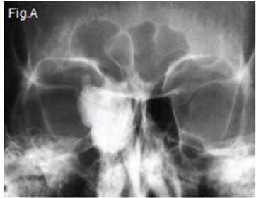
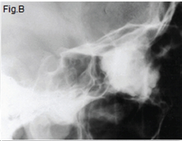
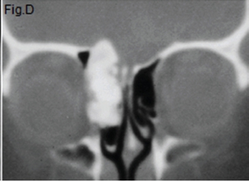
Upper left (Fig. A) Occipital mental view (caudally angled by 15 degrees) and upper right lateral skull view (Fig. B) shows a large, dense mass occupying the right ethmoid region and expanding into the right orbit. The lower right axial CT (Fig. C) and lower left coronal CT (Fig. D) shows a mass of uniform density in the right ethmoid sinus region. A histological diagnosis of a compact osteoma of the right ethmoid sinus was made.

The osteoma is a slow growing benign bone tumour. It varies in size from a pea to a hen's egg and may fill the entire sinus and extend into neighbouring structures. It has an incidence of approximately 0.25% in all routine examinations of the paranasal sinuses. The frontal sinus is the most common site of occurrence and the tumour is found less frequently in the ethmoid cells. Osteomas rarely occur in the maxillary or sphenoid sinuses. Pathologically, osteomas may be divided into compact or hard and cancellous or soft varieties depending upon the constituents of the lesion. The cancellous osteoma probably represents a form of fibrous dysplasia and should not be considered a true bone neoplasm. The lesion is more common in males than in females. It most often occurs during puberty and in the second and third decades of life. Most of the patients are asymptomatic and the tumour is a coincidental discovery during radiographic examination. Osteoma of the frontal sinus may produce deformity of the forehead, involvement of the orbit, and extension of the anterior cranial fossa. The radiological features of the compact osteoma are sharply defined, homogeneous rounded or lobulated, ivory-hard bony mass, either sessile or pedunculated. This type of osteoma is common. The cancellous osteoma appears as an irregularly defined, rounded or lobulated mass of less density, and may be mistaken for a soft tissue mass. This type of osteoma is rare. Gardner syndrome and familial adenomatous polyposis may be associated with multiple osteomas.
Reference
1. Dodd GD, Jing B: Radiology of the Nose, Paranasal Sinuses and Nasopharynx, Williams & Williams, Section 2 1977 p 180-181 [ Links ]
