


Services on Demand
Article
 English (pdf)
 Article in xml format
 Article references
Indicators

Related links
-
 Cited by Google
 -
 Similars in Google
Share

 Permalink
South African Dental Journal
On-line version ISSN 0375-1562
Print version ISSN 0011-8516
S. Afr. dent. j. vol.71 n.2 Johannesburg Mar. 2016
FORENSIC CASE
Forensic dentistry case book 6: A self-inflicted bite mark; a case report
S MaistryI; V M PhillipsII
IBSc; BSc Hons; MBCHB, Dip For. Med (SA); FC For. Path (SA); MMED For. Path (UCT). Department of Forensic Medicine and Toxicology. University of Cape Town
IIBDS; MChD; Dip. Max-Facial Radiol; FC Path SA (Oral Path); PhD; D.Sc. Department of Forensic Medicine and Toxicology. University of Cape Town
CASE REPORT
The case presented is of a 35 year old woman who was a victim of a vicious assault. At autopsy there were 26 stab wounds to the body, numerous defence injuries, her throat was slit and there was generalised organ pallor as a result of exsanguination. The stab wounds fractured the cervical spine and injured the lungs, oesophagus, trachea as well as the arteries and veins of the neck. On the left arm were superficial lesions consistent with a bite mark (Figure 1). The bite mark was photographed and swabbed for DNA. An impression of the bite mark was not taken because the lesions were very superficial and there was little penetration of the skin.

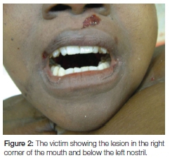
The examination of the victim's mouth showed a small red abrasion present on the upper lip near the right corner of the mouth (Figure 2). This abrasion was adjacent to the upper right canine, and was possibly caused by forceful pressure of the lip against the underlying canine. There was also an abrasion of the upper lip below the left nostril; that possibly occurred during the attack on the victim. Examination of the teeth of the victim showed no abnormalities.
The bite mark on the arm showed that there were two distinct curved patterns of lesions (Figure 1) indicating that there were two bite marks in this area.
Photographs were taken of the bite marks using an ABFO No. 2 mm scale (American Board of Forensic Odontology) to reference the size of the lesions produced by the teeth that had caused the pattern of bruises and abrasions. For analytical purposes the photographs were printed to the original size of the bite marks on the arm; i.e. 1:1 ratio.
When a bite mark occurs on the lower arm it is important to consider self-infliction as a possibility. The arm or hand is often forced into the victim's mouth to minimize minimise screaming. In this case, when the left arm was placed adjacent to the mouth it showed that there was a distinct possibility that the victim had bitten her own arm. At this stage of the investigation no suspect had been arrested.
The analysis of the bite mark continued with the comparison of the victim's teeth with the bite mark. Dental impressions of the victim's teeth were taken using President® silicone impression material. From these dental impressions upper and lower plaster of Paris study models were cast of the teeth for comparison purposes. These were used to duplicate the biting patterns of the upper and lower teeth of the victim by pressing the teeth of the study models into softened grey dental bite-registration wax. This resulted in an accurate bite pattern for the upper and lower teeth which was then compared with the actual bite mark. The patterns of the teeth in the wax are traced onto plastic foil with a permanent marking pen and then super-imposed over the bite mark to elicit concordant features (Figure 3).

The illustrations in Figure 3 demonstrate the relationship between the victim's upper teeth and the bite mark on the left arm. The dental arch matched the outer pattern of bruises.
The lower illustration shows the tracing of the biting pattern of the victim's upper teeth superimposed on the photograph of the bite mark. Six concordant features were identified.
RESULTS
The analysis of the bite marks in comparison with the teeth of the victim showed the following:
- Six concordant features between the upper teeth and the upper outer aspect of the bite mark
- Seven concordant features between the upper teeth and the upper inner aspect of the bite mark
- Ten concordant features between the lower teeth and the lower area of the bite mark.
DISCUSSION
IForensic pathologists need to be aware at autopsy that bruises and abrasions could possibly be a bite mark.1 There are several important lessons from this case study.
Firstly, the time lapse between the infliction of a bite mark and when it is examined is critical.2-4 If a bite mark is analysed soon after the event the chances of making an accurate match with the perpetrator's, or in this case, the victim's, teeth, are greatly improved. Secondly the skin and underlying tissues of the forearm are relatively soft and malleable and allow a degree of distortion when bitten. Therefore the abrasions and bruises may be somewhat mis-shaped when compared with the teeth and the biting patterns of whoever inflicted the bite.5,6
The ideal analysis of a bite mark is to take an impression of the puncture wounds and to cast a model; this creates an accurate replica of the bite mark which can be compared with the perpetrator's teeth. In this case the bruises and abrasions on the arm were too superficial to justify an impression.
Photographs of the bite marks need to be printed to as close to the original size of the bite mark seen on the victim i.e. 1:1 ratio for accurate analysis. In this case, the clinical and photographic examination showed distinct crescent shaped patterns of bruises and abrasions on the skin, caused by the upper and lower teeth. The bite marks were on the inner aspect of the left forearm of the victim. This suggested that there was a distinct possibility that the arm of the victim was forced into her mouth during the attack and that the bite mark was self-inflicted.10-12
The arms are usually raised during an attack with the outer surface towards the attacker, therefore a bite inflicted by the attacker would be on the outer surface of the arm.7,8 The abrasion and bruise patterns also suggest a struggle because there were abrasions on the skin produced by the upper teeth that appear to have been dragged across the skin surface due to the victim trying to remove her arm from her mouth. The right corner of the victim's mouth shows a lesion on the upper lip that may have been caused by the victim's right canine during forceful insertion of her left forearm into her mouth. The bite marks in this victim were mostly abrasions with no puncture wounds, further supporting self-infliction.12 A bite mark by an attacker is usually more severe with bleeding puncture wounds.8,13
There is no consensus as to the minimum number of concordant features in bite mark analysis which are necessary to determine a complete match between the teeth of the perpetrator (or victim) and a bite mark.9 The greater the number of concordant features, the higher the probability of a match. Hence in this case the probability that the victim bit her own arm is high.13
CONCLUSION
Bite marks are frequently encountered by Forensic Pathologists at autopsy. However, analysis of a bite mark requires the expertise of a Forensic Odontologist. This case report has demonstrated the methods used to analyse a bite mark and the need for Forensic Pathologists to be aware of bite marks and to take DNA swabs before the autopsy. The number of concordant features in this case suggested that there was a high degree of probability that the bite mark on the left forearm of the victim was self-inflicted.
ACRONYM
ABFO: American Board of Forensic Odontology
References
1. Saukko P, Knight B. Knight's Forensic Pathology. 3rd Ed. Hodder Arnold. 2004; Chap. 26 pg.527-41 [ Links ]
2. Thompson, I.O.C., Phillips, V.M. Bite mark case with a twist. The Journal of Forensic Odonto-Stomatology. 1994; 12: 37-40. [ Links ]
3. Lessig R, Wenzel V, Weber M. Bite mark analysis in forensic routine case work. EXCLI Journal. 2006; 5: 93-102. [ Links ]
4. Dorian RBJ. Human Bite marks in: Bite-mark Evidence: A Colour Atlas and Text, 2nd Ed. Florida: CRC press. 2011; Ch.18, pg. 269-70. [ Links ]
5. Sheasby DR & MacDonald DGA. Forensic classification of distortion of human bite marks. Forensic Science International. 2001; 122 (1): 75-8. [ Links ]
6. DeVore DT. Bite marks for identification? A preliminary report. Medicine Science and Law.1971; 11 (3): 144-5. [ Links ]
7. Vale GL and Noguchi TT. Anatomical distribution of human bite marks in a series of 67 cases. Journal of Forensic Sciences. 1983; 28 (1): 61-9. [ Links ]
8. Petty IA and Sweet D. Anatomical location of bitemarks and associated findings in 101 cases from the United States. Journal of Forensic Sciences. 2000;45 (4): 812-4. [ Links ]
9. Bowers CM and Pretty IA. Expert disagreement in bite mark casework. Journal of Forensic Sciences. 2009; 54 (4): 915-8. [ Links ]
10. Sobel MN and Perper JA. Self-inflicted bite mark on the breast of a suicide victim. American Journal of Forensic Medicine and Pathology.1985; 6 (4): 336-8. [ Links ]
11. Warnick AJ, Biedrzycki L, Russanow G. Not all bite marks are associated with abuse, sexual activities, or homicides: a case study of a self-inflicted bite mark. Journal of Forensic Sciences. 1987; 32 (3): 788-92. [ Links ]
12. Ronchese, F. Self-inflicted bite mark. American Journal of Surgery. 1994; 6 (1): 80-5. [ Links ]
13. Furness J. A general review of bite mark evidence. American Journal of Forensic Medicine and Pathology.1981; 2 (1): 49-52. [ Links ]
 Correspondence:
Vincent Michael Phillips
Department of Oral and Maxillofacial Pathology & Forensic Sciences
Faculty of Health Sciences, University of the Western Cape
South Africa
Tel: 021 937 3161
E-mail: vmphillips@uwc.ac.za
