


Services on Demand
Article
 English (pdf)
 Article in xml format
 Article references
Indicators

Related links
-
 Cited by Google
 -
 Similars in Google
Share

 Permalink
South African Dental Journal
On-line version ISSN 0375-1562
Print version ISSN 0011-8516
S. Afr. dent. j. vol.70 n.9 Johannesburg Oct. 2015
RADIOLOGY CASE
Maxillo-facial radiology case 136
CJ Nortjé
BChD, PhD, ABOMR, DSc. Faculty of Dentistry, University of the Western Cape. E-mail: cnortje@uwc.ac.za
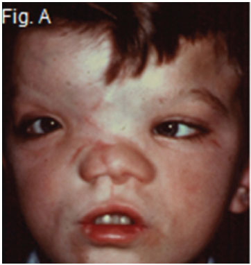
Below are a clinical picture and skull radiographs of a patient having a developmental field defect, probably occurring between 21 and 70 days of uterine life, rather than an individual syndrome. As such the etiology and pathogenesis are probably heterogeneous. What is your diagnosis?
INTERPRETATION
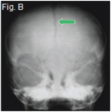
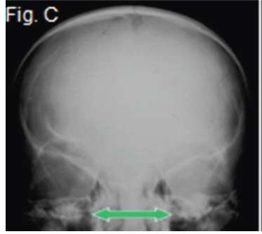
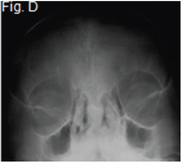
A diagnosis of frontonasal malformation was made. Frontonasal malformation has been defined as a combination of two or more of the following characteristics: hypertelorism, broadened nasal bridge, medium facial cleft affecting the nose and the upper lip and sometimes the palate, unilateral or bilateral clefting of the nasal alae, lack of formation of the nasal tip. The appearance of cranium bifidum (also known as cleft skull or enlarged parietal foramina) is characterized by the unsuccessful midline migration of the cranial vault, and a V-shaped hairline prolongation onto the middle of the forehead. The clinical picture (Fig. A) shows many of the characteristics mentioned above. The postero-anterior view of frontonasal malformation (Fig.B) shows hypertelorism, a widened nasal bridge and persistence of the metopic suture (arrow). A further postero-anterior view (Fig. C) shows persistence of the anterior fontanelle and a widened nasal bridge (arrow) with concomitant hypertelorism. The Waters view (Fig.D) demonstrates the hypoplastic maxillary sinuses. There is also marked hypertelorism and a widened nasal bridge.
Reference
1. Farman AG, Nortjé CJ, Wood RE. Oral and Maxillofacial Imaging, 1st Ed, Mosby, St. Louis, Missouri, 1993, pp. 122-123 [ Links ]
