


Services on Demand
Article
 English (pdf)
 Article in xml format
 Article references
Indicators

Related links
-
 Cited by Google
 -
 Similars in Google
Share

 Permalink
South African Dental Journal
On-line version ISSN 0375-1562
Print version ISSN 0011-8516
S. Afr. dent. j. vol.70 n.8 Johannesburg 2015
RADIOLOGY CASE
Maxillo-facial radiology case 135
CJ Nortjé
BChD, PhD, ABOMR, DSc. Faculty of Dentistry, University of the western Cape. e-mail: cnortje@uwc.ac.za
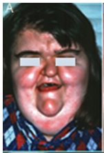
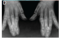
This patient presented with a sunken concave midface, anterior open bite, vertically elongated head, wide spread bulging eyes, various impacted teeth and chronic pain on the left side of the face. She also has syndactyly of the hands and feet. The same condition is also present in several members of her family. What is your diagnosis?
INTERPRETATION
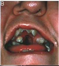
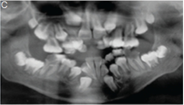
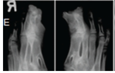
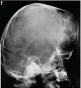
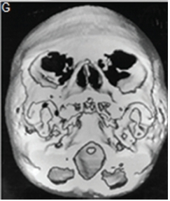
A diagnosis of Apert syndrome was made. Apert syndrome is a rare developmental condition characterized by premature cranial synostosis and resultant growth disturbances. Signs of Apert syndrome include a peaked and vertically elongated head, widespread bulging eyes, and a protuberant frontal region with an anteroposterior ridge overhanging the frontal eminence (Figure A). The palate is high, arched and occasionally cleft (Figure B). There is hypoplasia of the maxilla with relative prognatism of the mandible. Dental malocclusions with crowding and delayed dental eruption in the maxilla are common (Figure C). The facial angle is exaggerated; the nose is small and has been compared to a parrot's beak in appearance. Hypertelorism, exophthalmos and divergent strabismus are often present; sometimes with blindness. Spina bifida has been recorded in some patients. Syndactyly of the hands and feet varies greatly. Hand films in Apert syndrome showing syndactyly with fusion of three fingers in both hands and webbing (Figure D). Syndactyly of the feet in the same patient is also discernible (Figure E) Note the tall (turricephalic) skull, open metopic suture, and faint beaten-silver appearance of the calvarium. Skull base and roof of the calvarium are flattened, with a noticeable beaten-silver appearance (Figure F). The 3-D CT reconstruction of the same patient showing hypoplastic maxilla with posterior cleft (Figure G) The patient may be retarded or of normal intelligence. Apert syndrome may be associated with advanced paternal age. The cardinal radiologic features of Apert syndrome are: Brachycephalic (reduced anteroposterior dimension of the skull with increased skull width). Turricephaly (occurrence of a skull with high vertical index), beaten silver appearance of the calvarium, absence of demonstrable cranial sutures in coronal dimension in young patients, hypoplastic maxilla and syndactyly of the hands and feet. Differential Diagnosis: Crouzon's disease, Pfeifer syndrome, Carpenter syndrome and Summit syndrome.
Reference
1. Farman AG, Nortje CJ, Wood RE. Oral and Maxillofacial Imaging, 1st Ed, Mosby, St. Louis, Missouri, 1993, pp. 107-109. [ Links ]
