


Serviços Personalizados
Artigo
 Inglês (pdf)
 Artigo em XML
 Referências do artigo
Indicadores

Links relacionados
-
 Citado por Google
 -
 Similares em Google
Compartilhar

 Permalink
South African Dental Journal
versão On-line ISSN 0375-1562
versão impressa ISSN 0011-8516
S. Afr. dent. j. vol.70 no.3 Johannesburg Abr. 2015
RADIOLOGY CASE
Maxillo-facial radiology case 129
CJ Nortjé
BChD, PhD, ABOMR, DSc. Faculty of Dentistry, University of the western Cape. e-mail: cnortje@uwc.ac.za
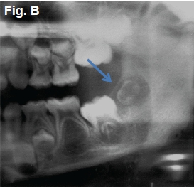
This six-year-old female patient (Figure A) presented with episodes of oral bleeding, fever and ulcerations affecting the submandibular region. Cropped pantomographs of the same patient were taken six weeks earlier (Figure B) with a follow-up pantomograph six weeks later (Figure C). What are the most important radiological differences between the cropped pantomographs?


INTERPRETATION
The important differences between the cropped panto-mographs are the displacement of the 2nddeveloping permanent molar (blue arrows) and loss of bone surrounding the mandibular primary 2nd molar (red arrow). A diagnosis of acute leukemia was made. Acute leukemia occurs mostly during the first decade of life, usually with gradual onset followed by a rapid course of a few weeks to a few months. In general, it is difficult to distinguish between the various types of acute leukemia's as the cells are in an immature state. Oral manifestations are often found in the form of gingival abnormalities, petechiae and ecchymosis, ulcers and bleeding of decreasing frequency. Very frequent is lymphadenopathy of the submandibular region. Radiological changes are said to occur in 60 % of children and 10% of adults. In adults changes are preferentially seen in the axial skeleton; in children the appendicular skeleton is most commonly involved. Overall 50% of patients show radiological changes and 63% exhibit abnormalities on panoramic radiographs. The cardinal radiological signs of acute leukemia affecting tha jaws are: premature loss of teeth, generalized absence of the lamina dura (yellow arrow) (Figure D), resorption of bone around the roots of both mandibular and maxillary first molar teeth, (green arrows) (Figure E), periapical radiolucency simulating typical inflamatory periapical changes, single, non cyst-like radiolucency of the alveolar bone and generalized rarefaction of the jawbones, which may take the form of a reduction in the number of radiographically demonstrable trabeculae in the mandible or maxilla (Figure E). Ancillary radiological signs of acute leukemia are: absence of the follical walls of unerupted teeth, multiple well-defined radiolucencies, teeth including those in crypts displaced. Differential diagnosis inlude: hyperparathyroidism, neuroblastoma metastatic to bone and renal osteodystrophy.


Reference
1. Pindborg, J.J &Hjorting-Hansen, E: Atlas of Diseases of the Jaws. W.B Saunders, 1974: p 60-61. [ Links ]
2. Farman AG, Nortjé CJ & Wood R E: Oral and Maxillofacial Imaging, 1st Ed, Mosby. St. Louis, Missouri 1993 p 294-295. [ Links ]
