


Services on Demand
Article
 English (pdf)
 Article in xml format
 Article references
Indicators

Related links
-
 Cited by Google
 -
 Similars in Google
Share

 Permalink
South African Dental Journal
On-line version ISSN 0375-1562
Print version ISSN 0011-8516
S. Afr. dent. j. vol.70 n.1 Johannesburg Feb. 2015
RADIOLOGY CASE
Maxillo-facial radiology case 127
CJ Nortjé
BChD, PhD, ABOMR, DSc. Faculty of Dentistry, University of the Western Cape. E-mail: cnortje@uwc.ac.za
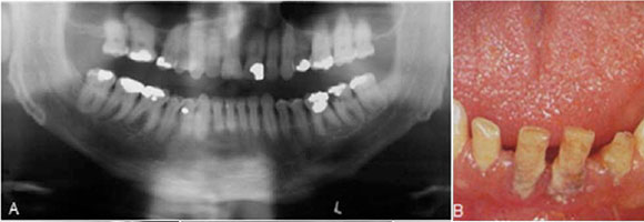
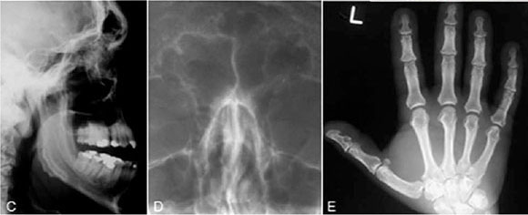
Below are clinical and radiographic images of a condition in which the terminal phalanges of the hands and the feet are enlarged. Growth of the mandibular condyle results in marked prognathism, and this together with macroglossia, leads to dental malocclusion. Which radiograph points most directly to the diagnosis?
INTERPRETATION
All the features mentioned above as well as the enlarged pituitary fossa/sella tursica (Figure C) is suggestive of acromeg aly. Hyperpituitarism is generally idiopathic in children and is caused by an acidophilic adenoma of the anterior lobe of the pituitary gland in adults. In childhood, hyperpituitarism leads to gigantism; in adulthood it causes acromegaly. Both are caused by an increased secretion of growth hormone stemming from an eosinophylic hyperplasia or in most cases as eosinophylic adenoma of the anterior pituitary gland. The disorder is most frequently seen between the ages of 20 and 40 years, but onset of the condition is often in late adolescence. Males and females are equally affected. When hyperpituitarism occurs after epiphyseal closure, acromegaly results. The terminal phalanges of the hands (Figure E) and of the feet are enlarged, and the ribs are increased in size. Growth of the mandibular condyles and rami results in marked prognathism, and this together with macroglossia (Figure B) leads to dental malocclusion. Spreading of the teeth, especially of the mandibular incisors, is a most characteristic change and is often an early symptom. The enlarged tongue shows indentations on the sides because of pressure against the teeth. The skin is coarse, and the lips become thickened. Headaches, photophobia, and reduced vision may also be present. Radiographically, one sees most frequently an enlarged sella tursica (Figure C) and a dramatic increase in size of the frontal sinuses (Figure D). Apart from the relatively frequent mandibular protrusion, a flattening of the gonial angle (Figure A) is often seen in acromegaly. Resorption of the roots as well as excessive hypercementosis has been reported. Therapeutically, the adenoma of the pituitary gland is treated surgically or by radiotherapy.
Reference
1. Pindborg, J.J & Hjorting-Hansen, E: Atlas of Diseases of the Jaws. W.B Saunders, 1974: p 118-9. [ Links ]
2. Farman AG, Nortjé CJ & Wood R E: Oral and Maxillofacial Imaging, 1st Ed, Mosby. St. Louis, Missouri 1993 p 331-2 [ Links ]
