


Services on Demand
Article
 English (pdf)
 Article in xml format
 Article references
Indicators

Related links
-
 Cited by Google
 -
 Similars in Google
Share

 Permalink
South African Dental Journal
On-line version ISSN 0375-1562
Print version ISSN 0011-8516
S. Afr. dent. j. vol.69 n.10 Johannesburg Nov. 2014
RADIOLOGY CASE
Maxillo-facial radiology case 126
CJ Nortjé
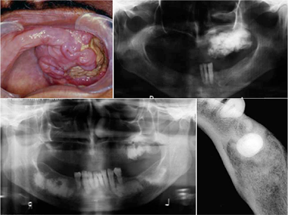
A fifty year old black female patient presented with the main complaint that a painful swelling in the upper left maxilla had been growing slowly over the last nine months (upper clinical and cropped pantomograph). Clinical examination revealed an ulcer-ative lesion showing expansion of the jaw.
INTERPRETATION
The upper clinical picture shows an expansive lesion and the cropped pantomograph, a diffuse sclerotic mass in the left maxilla. Provisional diagnosis of florid cement-osseous dysplasia (also diagnosed as gigantiform cementoma by some pathologists some years ago) was made, which was confirmed histopathologically. Florid cement-osseous dysplasia is a progressive patchy sclerosis of the jaws found predominantly in black women aged 25 years and older. The disease is idiopathic in most cases, though a familial pattern is sometimes present. Close to 90% of cases involve the mandible, and nearly 60% involve all four quadrants (lower right pan-tomograph) and the premolar region is the most commonly affected.1 The lower mandibular occlusal radiograph reveals patchy sclerosis in the premolar region, somewhat expanding the jaw, however, the sclerotic masses do not reach the mandibular cortex. Radiographically, the lesion presents as dense radiopaque masses which sometimes are distributed more or less symmetrically in the jaws, thus supporting the view that they represent some form of dysplasia or developmental anomaly. Other cardinal radiographic features include a pagetoid cotton wool appearance, a thin radiolucent band at the periphery of the sclerotic masses and a "ground glass" appearance of the alveolar bone with loss of the periodontal ligament spaces. Florid cement-osseous is usually asymptomatic unless secondary osteomyelitis or chronic diffuse osteomyelitis is superimposed. The condition is generally found coincidentally during radiographic examination of the jaws. As the lesion may occur in several members of a family it has been called familial multiple cementomas. Florid cement-osseous dysplasia requires treatment only if the lesion becomes disfiguring or is the site of a secondary infection, usually as an osteomyelitis.
Reference
1. Farman AG, Nortjé CJ & Wood R E: Oral and Maxillofacial Imaging, 1st Ed, Mosby. St. Louis, Missouri 1993 p 319-323. [ Links ]
