


Services on Demand
Article
 English (pdf)
 Article in xml format
 Article references
Indicators

Related links
-
 Cited by Google
 -
 Similars in Google
Share

 Permalink
South African Dental Journal
On-line version ISSN 0375-1562
Print version ISSN 0011-8516
S. Afr. dent. j. vol.69 n.9 Johannesburg Oct. 2014
RADIOLOGY CASE
Maxillo-facial radiology case 125
CJ Nortjé
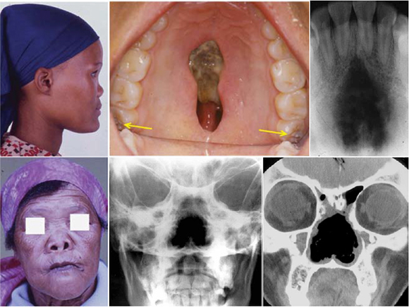
Below are clinical and radiological pictures of two patients with the same pathology. The upper case Is that of a 20-year old who has been aware of a palatal lesion since birth and the lower case is that of a 50-year old who presented with destruction of the nasal septum and conchae.
INTERPRETATION
The 20 year old woman was referred to the Faculty for investigation of a large palatal defect. She had been diagnosed previously as having congenital syphilis. At her initial presentation it was found that she had a positive Venereal Research Laboratory (VDRL) test, a negative fluorescent treponemal antibody absorption test (FTA-ABS) and a negative T. pallidum haem-agglutination test. Physical examination revealed erosion of the nasal septum with a large gumma evident in the palate. The clinical pictures show a depression of the nasal bridge and a large gumma in the midline of the palate. The occlusal radiograph of the area reveals a poorly demarcated radiolucency in the maxillary midline. The 18 and 28 (yellow arrows) show the characteristic enamel hypoplasia of congenital syphilis ("mulberry molars"). A diagnosis of congenital syphilis was made. In the lower case the clinical picture shows a gummatous destruction of the bridge of the nose. The middle posterior-anterior view shows gummatous destruction of the nasal septum and conchae which are also discernible on the lower right coronal CT view. A diagnosis of a tertiary stage of syphilis was made. Syphilis is caused by infection by the spirochete Treponema pallidum and may be congenital or acquired. The acquired form is usually further sub-classified into three distinct stages: primary, secondary and tertiary. The bone may be affected in congenital syphilis and in both secondary and tertiary stages of acquired syphilis. The jaws are rarely affected in syphilis. When they are, the palate is more frequently involved than the mandible. The characteristic lesion of primary syphilis is the chancre, which clinically presents as an ulcerated, indurated, painless lesion. Oral lesions of secondary syphilis include mucous patches and maculopapular eruptions. Two types of lesions occur in tertiary syphilis, the gumma and atrophic syphilitic glossitis. The cardinal radiologic features of syphilis are deposition of sub- periosteal new bone along the inferior border of the mandible (syphilitic periostitis). Destruction of bone, especially the palate, causes perforation of the palate and appears as large radiolucent areas (gumma). Well demarcated destruction is noted along cortical margins, especially the mandible (cortical gummas). Multiple radiolucencies with poorly defined margins and sequestration formation (syphilitic osteomyelitis), are observed.
Reference
1. Farman AG, Nortjé CJ & Wood R E: Oral and Maxillofacial Imaging, 1st Ed, Mosby. St. Louis, Missouri 1993 p 204-5. [ Links ]
