


Services on Demand
Article
 English (pdf)
 Article in xml format
 Article references
Indicators

Related links
-
 Cited by Google
 -
 Similars in Google
Share

 Permalink
South African Dental Journal
On-line version ISSN 0375-1562
Print version ISSN 0011-8516
S. Afr. dent. j. vol.69 n.8 Johannesburg 2014
RADIOLOGY CASE
Maxillo-facial radiology case 124
CJ Nortjé
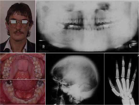
A healthy 24-year-old male presented at the Faculty complaining of painful headaches, back pain and a jaw deformity: his chin and lower jaw had become so enlarged that the chin strap on his motorcycle helmet no longer fitted. He reported he had been well until seven years previously when he began to experience painful bilateral temporal headaches: five years later he had a transient paralysis of the left facial nerve and six weeks ago he had spontaneous lacrimation of the left eye and dilation of the left pupil. Physical examination revealed a healthy tall adult male with large facial features but clinically normal hands: there was marked increase in the height of the mandible (pantomograph). Intra-orally, there were lobulated tori of the hard palate and lingual tori in the premolar region (lower clinical pictures).
INTERPRETATION
A provisional diagnosis of osteopetrosis with mandibular prognathism and facial nerve paralysis was made. The skull radiographs showed increased thickness of the vault, especially in the occipital bone and base (lower skull radiograph). Carpal radiographs revealed involvement of the bones of the hand showing increased girth of the diaphyses and relative thickening of the phalangeal cortices, while the metacarpals and phalanges were cigar-shaped. The remainder of the skeleton exhibited a generalized increased density. These new findings were more consistent with a diagnosis of scleros-teosis rather than osteopetrosis and the patient was therefore referred to the neurosurgical services. Sclerosteosis is one of a group of disorders known as sclerosing bone dysplasias1 The condition was first described by Truswell2 but the term sclerosteosis is attributed to Hansen.3 It is most common in the Afrikaner population of South Africa but is otherwise considered rare. It is inherited as an outosomal recessive, in contrast to benign osteopetrosis which is dominant and is similar to another sclerosing bone dysplasia, van Buchem's disease, in that both are recessive, exhibit the radiographic features of osteopetrosis and are associated with jaw enlargement. It is vital that the condition be differentiated from osteopetrosis since sclerosteosis may be associated with sudden death secondary to impaction of the medulla oblongata due to elevated intracranial pressure.4
Reference
1. Beighton P, Davidson, Durr L and Hammersma H: The clinical features of sclerosteosis. A review of the manifestations in twenty-five affected individuals. Ann Int Med 84, 1976 :393-7. [ Links ]
2. Truswell, A.S.: Osteopetrosis with Syndactyly, a morphologic variation of Albers-Schonberg disease. J Bone Jt Surg 1958;40 (b):208-18. [ Links ]
3. Hansen, H, Sklerosteose G. In: Opitz, H, and Schmidt F. Handbuch der Kinderheilkunde, 6: Berlin, Springer-Verlag, 1967, pp.351-5. [ Links ]
4. Beighton, P and Cremin, BJ: Sclerosing Bone Dysplasias, Berlin, Springer-Verlag, 1980, pp19-188. [ Links ]
