


Services on Demand
Article
 English (pdf)
 Article in xml format
 Article references
Indicators

Related links
-
 Cited by Google
 -
 Similars in Google
Share

 Permalink
South African Dental Journal
On-line version ISSN 0375-1562
Print version ISSN 0011-8516
S. Afr. dent. j. vol.69 n.8 Johannesburg 2014
CLINICAL COMMUNICATION
Radix Entomolaris - a rare case series
S MahalakshmiI; M Archana ShenoyII; B ShubhaIII; CJ ChandyIV
IMDS. Senior Lecturer, Department of Oral Medicine and Radiology, Sri Siddhartha Dental College & Hospital, Sri Siddhartha Academy of Higher Education, Tumkur, Karnataka State, India
IIBDS. Tutor, Department of Conservative Dentistry and Endodontics, Sri Siddhartha Dental College, Agalakote, Tumkur, Karnataka State, India
IIIBDS. Tutor, Department of Pedodontics, Sri Siddhartha Dental College, Agalakote, Tumkur, Karnataka State, India
IVSr. Lecturer, Department of Oral Medicine and Radiology, Mar Baselios Dental College, Kothamangalam, Kerala state, India
ABSTRACT
INTRODUCTION: The success of root canal treatment is determined by satisfying the basic principles of complete removal of the endodontic pathology through chemo-mechanical cleansing of the root canals followed by shaping and then by obturation to provide the ideal hermetic seal. To achieve such success a thorough knowledge of the root canal anatomy is a basic requirement. Several anatomic variations of the mandibular molars have been reported among which is the reportedly rare Radix Entomolaris (RE), a mandibular molar with an additional root located lingually.
AIMS AND OBJECTIVES: To present five cases of RE diagnosed pre-operatively using radiographs and subsequently successfully endodontically treated. We also aim to highlight the considerations for the diagnosis and management of RE in general dental practice.
CONCLUSION: Even though RE is rarely encountered in general dental practice, the possibility that it may occur warrants the need to have sufficient knowledge regarding diagnosis and the appropriate modifications in endodontic management of these teeth. Preparedness coupled with a carefully modified clinical approach aids in successful management of RE and ensures that these cases could be handled with ease.
Key words: Endodontic treatment, mandibular molar, anatomical variations, radix entomolaris.
ACRONYMS
ALARA: As low as reasonably achievable
CBCT: Cone Beam Computed Tomography
CT: Computed Tomography
PDL: Periodontal Ligament
RCT: Root Canal Treatment
RE: Radix Entomolaris
RP: Radix Paramolaris
INTRODUCTION
The success of root canal treatment is determined by satisfying the basic principles of complete removal of the endodontic pathology through chemo-mechanical cleansing of the root canals, followed by shaping and then by obturation to provide the ideal hermetic fluid-tight seal.1 The primary aim of endodontic treatment is the elimination of bacteria from the infected root canal and the prevention of subsequent reinfection.2 To achieve this, a thorough knowledge of the variations in root canal anatomy is absolutely imperative as diversities such as extra roots, extra canals, webs, fins, and isthmuses may complicate the endodontic procedure.2 The majority of mandibular first molars are two-rooted (97,8%) with three canals.3,4 The mesial root usually has two canals ending in two distinct apical foramina or they may merge to end in a single foramen. The distal root most commonly has one large kidney-shaped canal, but if the orifice is narrow and round, a second canal may be present1 The incidence of occurrence of two distal canals is reported to be 28%.5 Several anatomic variations of the mandibular molars have been reported among which are, notably, Radix Entomolaris(RE), a mandibular molar with an additional root located lingually, and Radix Paramolaris(RP) with the additional root located buccally.6
RE was first reported by Carabelli in the year 1844.7,8 A classification by Carlsen and Alexandersen24 describes four different types of RE according to the location of the cervical part of the RE: types A, B, C and AC. Types A and B refer to a distally located cervical part of the RE with, respectively, two normal and one normal distal root components. Type C refers to a mesially located cervical part, while type AC refers to a central location, between the distal and mesial root components. This classification allows for the identification of separate and non-separate RE.1 Another classification is that of De Moor et al,7 which is based on variations in the bucco-lingual curvature of the root canal. Three types can be identified: Type I refers to a straight root/root canal, while Type II refers to an initially curved entrance which continues as a straight root/root canal. Type III is characterized by having an initial curve in the coronal third of the root canal and a second curve be ginning in the middle and continuing to the apical third. The frequency of RE is less than 5% in white Caucasian (UK, Dutch, Finnish, German), African (Bantu, San), Eurasian and Indian populations. Certain ethnic populations with Mongoloid traits, such as the Chinese, Eskimos and Native American population show a frequency of 5% to 36%.8 This rare malformation may confront the general practitioner who has to manage an RE with pulpal involvement. The treatment may develop complications if a canal is missed during endodontic therapy or if the extra root fractures and may be retained during exodontia. Care during the initial radiographic evaluation should forewarn the general practitioner, who is then prepared and will handle these cases with ease. Radiographs taken at different angulations reveal the basic information regarding the anatomy of a tooth and can thus help in the detection of any aberrant anatomy such as extra canals/roots.10 If conventional radiography still leaves an element of doubt then advanced imaging with CBCT will reveal the three-dimension curvature and angulation of the canals and roots. The dentist can anticipate the difficulties and should now decide whether to refer the patient to an endo-dontist or to manage the patient in the general practice.11,12
This study presents five cases of RE diagnosed pre-oper-atively using radiographs and successfully treated with endodontic therapy.
CASE REPORTS
Two male and three female Indian patients reported at the Hospital complaining of symptoms suggestive of pulpal involvement of the RE. A detailed history was elicited following which clinical and radiological evaluation revealed that root canal treatment (RCT) was indicated in all five cases. RE were identified by assessment of the pre-operative radiographs. A systematic and sequential approach to treatment was adopted for all the cases. Initially, before treatment, radiographs of the contralateral molars were taken for all the five patients as literature documents a high incidence of bilateral presentation of RE.13 None of our cases showed bilateral involvement. After achieving adequate local anesthesia the teeth were isolated using a rubber dam. The access cavities were prepared in the conventional manner and modified from a triangular to trapezoidal outline so as to locate the fourth distolingual canal. The root canals were located with an endodontic probe (DG-16). The canals were then explored with K-File ISO 15 (Dentsply Maillefer). The working lengths were determined electronically using Propex and confirmed with radiographs. The root canals were then shaped with Protaper rotary instrument in the first three cases while conventional hand filing and shaping was done in the fourth and fifth cases. Following debridement, the canals were irrigated with 2.5% sodium hypochlorite solution. At the next appointment, master cone radiographs were obtained following which the canals of the first three cases were obturated with Protaper F-2 gutta-percha points while the last two cases were obturated with conventional gutta percha points. The access cavities were restored with Miracle Mix. All cases were successfully treated. The morphological diversity of each case is documented below.
Case 1
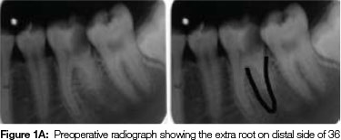
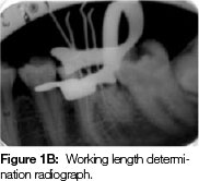
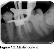
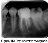
A 21 year old female patient presented with pain in her mandibular left first molar. Clinical examination revealed gross decay and tenderness on vertical percussion. Clinical and radiological evaluation revealed widening of the periodontal ligament (PDL) space and an increased radio-density surrounding the mesial root suggestive of a final diagnosis of apical periodontitis with condensing ostetitis in 36. The radiograph also revealed the presence of the extra root on the distal side of first molar (Figure 1A). The two mesial canal orifices were located. The distal canal orifice was slightly buccally away from the centre indicating the possibility of a disto-lingual canal. The disto-lingual root canal was 0.5mm more than the disto-buccal root. There was an initial curve in the coronal third of the canal and a second curve beginning in the middle and continuing to the apical third. (Figure 1B) This RE can be classified as Carlsen and Alexanderson TYPE -A and De Moor et at classification TYPE -III RE (Table 1). The irrigation, obturation and post obturation filling were completed as1 previously described. (Figure 1C, Figure 1D).
Case 2
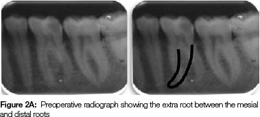
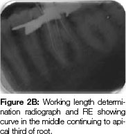
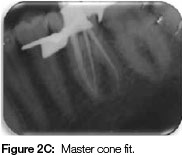
A 17 year old male patient presented with pain and swelling on the left side of the face of four days duration. It was a severe, continuous, throbbing type of pain that was increased on chewing food. On examination, the mandibular left first molar was decayed disto-proximally and the tooth was tender to percussion. The radiograph revealed no periapical changes but the extra root was clearly visible, located between the mesial and distal roots (Figure 2A). A final diagnosis of buccal space infection secondary to acute periapical abscess was made. In this case only an emergency access opening was performed at the first appointment to relieve the acute pain, following which antibiotics were prescribed. Coronal enlargement and debridement were undertaken at the next appointment. Insertion of the No. 15 file in the fourth canal showed a lingually oriented access inclination. Upon removal the tip was slightly deformed with a curvature to the mesial side at the position of the middle third of the root to the apex. The disto-lingual root canal was 3mm shorter than the disto-buccal (Figure 2B). As in the previous case this RE can also be classified as Carlsen and Alexanderson TYPE -A and De Moor et at classification TYPE -III RE. (Table 1) The irrigation, obturation and post obturation filling were finalized in the manner described above (Figure 2C, Figure 2D).
Case 3
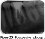
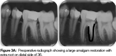
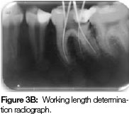
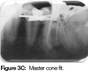
A 21 year old male patient presented with pain in his previously restored mandibular left first molar, which had a large amalgam restoration and was tender to percussion. The pre-operative radiograph revealed the filling in close proximity to the pulp, and a widening of the PDL space in the mesial root suggestive of apical periodontitis. The extra root could be discerned on the distal aspect as a superimposition with the middle and lower third appearing in between the mesial and distal roots (Figure 3A). The disto-lingual root was straight and the disto-lingual canal was easily located. It was 1mm shorter than the disto-buccalroot canal (Figure 3B). This RE can be classified as Carlsen and Alexanderson TYPE -A and De Moor et at classification TYPE -I RE (Table 1). The irrigation, obturation and post obturation filling were completed routinely (Figure 3C, Figure 3D).
Case 4
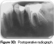
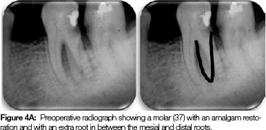
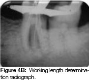
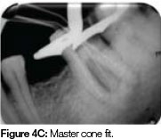
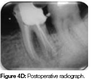
A 39 year old female patient presented with pain in her previously restored mandibular left second molar. Preoperative radiography showed a cervical radiolucency involving the pulp, a widening of PDL space around the mesial and distal roots, suggestive of apical periodontitis. The presence of the extra root on the distal side of the tooth was demonstrated (Figure 4A). The disto-lingual root canal was 3 mm shorter than the disto-buccal canal and was straight and easily negotiable (Figure 4B). This RE can be classified as Carlsen and Alexanderson TYPE -A and De Moor et at classification TYPE -I RE. (Table 1) The debridement, irrigation, obturation and post obturation filling were all completed satisfactorily (Figure 4C, Figure 4D).
Case 5
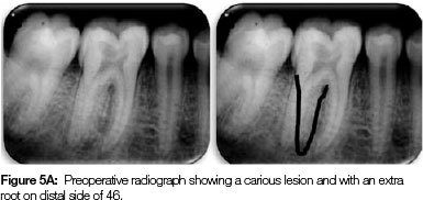
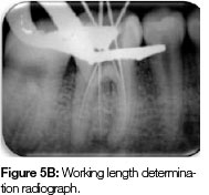
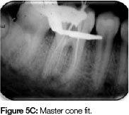
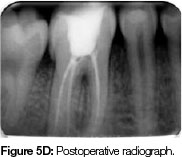
An 18 year old female patient reported with pain in her right mandibular first molar which on examination revealed a grossly decayed tooth with radiographic evidence of a widening of the PDL space suggestive of apical periodontitis. The unclear outline of the distal root contour gave rise to the suspicion of a superimposed extra root (Figure 5A). Another radiograph taken from a more mesial angulation clearly revealed the extra root. The disto-lingual root canal was 1 mm shorter than the dis-to-buccal canal and it was straight and easily negotiable (Figure5B). This RE can be classified as Carlsen and Alexanderson TYPE -A and De Moor et at classification TYPE -I RE. (Table 1) The debridement, irrigation, obturation and post obturation filling were done as mentioned earlier. (Figure 5C, Figure 5D).
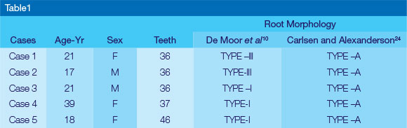
We have morphologically classified all the cases in our case series and these are documented in Table1.
DISCUSSION
Several anatomic variations of the mandibular molars6 have been reported among which are the rather unusual presentations: Radix Entomolaris(RE), a mandibular molar with an additional root located lingually and Radix Paramolaris(RP) with an additional root located buccally. The etiology behind the formation of RE is still unclear. Its formation could be related to external factors during odontogenesis.14 Another theory is that of an atavistic gene penetrance1 (Atavism is the reappearance of a trait after several generations of absence). In eumorphic roots, population specific factors influence the more profound expression of a particular gene that results in more pronounced phenotypic manifestations.15 Curzon suggested that the 'three-rooted molar' trait has a high degree of genetic penetrance as its dominance is reflected in the fact that the prevalence of the trait is similar in both pure Eskimo and Eskimo/Caucasian mixes. Third root anomalies may develop during bud morpho-differentiation as a result of a developmental aberration of both ectoderm and mesoderm.17
Various prevalence studies have been undertaken using periapical radiographs, extracted teeth and, more recently, micro computed tomography (micro-CT) and cone beam computed tomographic images (CBCT)18 In African populations a maximum frequency of 3%19 is found, while in Eurasian and Indian population the frequency is less than 5%. In populations with Mongoloid traits (such as the Chinese, Eskimo and American Indians) reports have noted that the RE occurs with a frequency that ranges from 5% to more than 30%. It has been speculated that recent studies will show higher prevalences compared with previous 2D image studies. This could possibly be a result of more accurate determination using 3D image analysis.18 In recent years, there has been an increasing number of case reports of re12,14,20,21 being published by Indian authors.2 However, this does not give any insight into whether the prevalence of RE in the Indian population is higher than previously documented and that could be the focus of future research.
RE is most commonly known to involve the mandibular first molar (7,4%) followed by the mandibular third molar (3.7%) involving least frequently the mandibular second molar (0.5%)1,22 Four of our cases involved the mandibular first molar while the fourth case involved the mandibular left second molar. According to Quackenbush, additional or 'extra' roots occurred unilaterally in approximately 40% of all cases and predominantly on the right side.23 Steelman observed this variation more frequently on the left side in Hispanic children.13 We recorded that four of our patients had RE on the left side while one involved the right side. Some studies have reported a bilateral occurrence of 50-67%.12'13This encouraged us to clinically and radiographically evaluate the contralateral molars for additional roots but none of our cases showed any bilateral involvement.
In general, the RE is smaller than the disto-buccal and mesial roots and can be separate from, or partially fused with, the other roots.10 In our case series, we observed that the disto-lingual canals of our second and fourth cases were 3mm shorter than the disto-buccal canal, while the working lengths were comparable in the other cases.
Subtle clinical features on intra-oral examination may point to the possibility of a supernumerary root. The presence of a more prominent disto-lingual lobe or an extra cusp in combination with a cervical prominence or convexity can indicate the presence of an additional root.1 However an increased number of cusps need not necessarily be associated with supernumerary roots. Hence identification of RE is not possible by clinical examination alone. Radiographic examination usually reveals the RE. In our case series, clinical examination did not contribute much to the identification of RE but the preoperative radiographs revealed their presence in all the five cases. Thorough inspection of the preoperative radiograph may show particular marks or characteristics such as an unclear outline of the distal/ mesial root contour or the root canal, probably because the supernumerary roots are mostly situated in the same bucco-lingual plane as the mesial or distal root resulting in superimposition.1 Sometimes on routine radiography the RE could be missed because of its slender morphology. A second radiograph of the tooth from a mesial or distal horizontal projection (20 degrees) may be helpful10 as would be specialized radiographic techniques like buccal object rule or "SLOB" technique. Advanced imaging modalities like micro-computed tomography18 and spiral computed tomography25 have been utilized for diagnosis of RE but we believe that resorting to these options does not adhere to the basic ALARA principle of diagnostic radiology. Cone beam computed tomography (CBCT) seems to be less invasive (an average of 52-1025pSv per exposure as compared with 2000-5000 pSv for spiral CT). The technique is accurate and has the additional advantage that the data is amenable to reformation in a volume rather than a slice, providing threedimensional images in the axial, coronal, or sagittal planes. CBCT is a promising tool for investigating the prevalence and morphologic feature of extra roots.26
Should preoperative radiographic evaluation fail to detect an RE, some clues observed during access cavity preparation may suggest the presence of an extra root or root canal. Typically the distal root has one kidney-shaped root canal. If the orifice is narrow and round, a second distal canal may be expected. A dark line on the pulp chamber floor can indicate the precise location of the RE canal orifice.1 When sodium hypochlorite is used in the pulp chamber, the remaining pulp tissue in the unidentified canal produces bubbles, a response which is referred to as the "champagne effect". This may also aid in locating the obscure canal.14 The identification of supernumerary roots and canals can be made easier with additional aids like the DG 16 probe, micro-opener, methylene blue dye, fluorescein sodium ophthalmic dye, long shank burs, ultrasonic instruments, surgical loupes, fibre-optic illumination, dental endoscopy and orascopy, operating microscope and visualization endogram using Ruddle's solution.18
Once a diagnosis of RE is made, the next step involves the modification of treatment strategy to suit the requirements of the RE. The orifice of the RE is located disto-to mesio lingually from the main canal.1 An extension of the original triangular opening cavity to the (disto-) lingual results in a more rectangular or trapezoidal outline form.10 If calcification is present above the orifice of the RE, this has to be removed to provide a better view. Care must be taken to not to remove an excessive amount of dentin on the lingual side of the cavity and orifice to avoid perforation of the coronal third of a severely curved root.18 Calberson et at1 recommend the use of flexible nickel-titanium rotary files to increase the chances of achieving a smooth glide path to the apical region. Nevertheless unforeseen complications can occur while negotiating the narrow canals with severe curvature.18
Apart from these endodontic implications, there are other considerations to be taken into account when dealing with RE in a general dental practice. During extraction of the RE, every effort should be made to avoid fracture of the curved root. An unidentified supernumerary root can fracture and even be retained following exodontia as the presence of the classical mesial and distal roots may lead the dentist to believe, mistakenly, that the tooth has been completely removed. Although the effect of RE on orthodontic treatment is not clear, it may be that the extra root increases the resistance to movement. The excessive cervical bulge in RE may be a contributing factor in localized periodontitis.
CONCLUSION
Clinicians should be aware of these unusual root morphologies in the mandibular first molars. Even though RE is rarely encountered, the possibility that it may occur warrants the need to have sufficient knowledge regarding diagnosis and the appropriate modifications in endodontic management of these teeth to prevent errors. Preparedness coupled with an appropriate and careful clinical approach aids in successful management of RE and ensures that these cases could be handled with ease.
Conflict of Interest: None declared
Author contributions: All the authors have contributed significantly towards the preparation of the preparation of final manuscript.
Acknowledgements: We would like to thank HOD and all the staff members from Department of Conservative and Endodontics for their support & cooperation.
References
1. Calberson FL, De Moor RJ, Deroose CA. The radix entomolaris and paramolaris: clinical approach in endodontics. J Endod 2007;33:58-63. [ Links ]
2. Attam K, Nawal R R, Utneja S, Talwar S. Radix entomolaris in mandibular first molars in Indian population: A review and case reports. Case Reports in Dentistry. 2012; Article ID 595494, 7 pages. [ Links ]
3. Barker BC, Parson KC, Mills PR, Williams GL. Anatomy of root canals. III. Permanent mandibular molars. Aust Dent J 1974;19:403-13. [ Links ]
4. Vertucci FJ. Root canal anatomy of the human permanent teeth. Oral Surg Oral Med Oral Pathol 1984;58:589 -99. [ Links ]
5. Pitt Ford TR. Pulp space anatomy and access cavities. In : Harty's Endodontics in Clinical Practice, 5th ed. Elsevier Science Limited, 2014:17-33. [ Links ]
6. Pineda F, Kuttler Y. Mesiodistal and buccolingual roentgeno-graphic investigation of 7,275 root canals. Oral Surg Oral Med Oral Pathol 1972; 33(1):101-10. [ Links ]
7. Carabelli G. Systematisches Handbuch der Zahnheilkunde, 2nd ed. Vienna: Braumuller und Seidel, 1844:114. [ Links ]
8. Bolk L. Bemerküngen über Wurzelvariationen am menschlichen unteren Molaren. Zeiting fur Morphologie und Anthropologie 1915;17:605-10. [ Links ]
9. Tratnman E K. Three-rooted lower molars in man and their racial distribution. Br. Dent J 1938; 64: 264-74. [ Links ]
10. De Moor R J, Deroose C A, Calberson F L. The radix entomolaris in mandibular first molars: an endodontic challenge. Int Endod J 2004;37:789 -99. [ Links ]
11. Cotton T P, Geisler T M, Holden D T, Schwartz S A, Schindler W G. Endodontic applications of cone-beam volumetric tomography. Journal of Endodontics 2007;33(9):1121-32. [ Links ]
12. Mahendra M, Verma A, Tyagi S,Singh S, Malviya K, Chaddha R. Management of complex root canal curvature of bilateral radix entomolaris: three-dimensional analysis with cone beam computed tomography. Case Reports in Dentistry 2013; Article ID 697323, 4 pages. [ Links ]
13. Steelman R. Incidence of an accessory distal root on mandibular first permanent molars in Hispanic children. J Dent Child 1986; 53:122-3. [ Links ]
14. Madhuram K, Keerthana S, Rajkumar S, Leena Sankari S. Radix Entomolaris: Report of two cases. Indian Journal of Multidisciplinary Dentistry 2011; 1 (4):227-30, [ Links ]
15. Reichart PA, Metah D. Three-rooted permanent mandibular first molars in the Thai. Community Dent Oral Epidemiol 1981; 9:191-2. [ Links ]
16. Curzon ME. Miscegenation and the prevalence of three-rooted mandibular first molars in the Baffin Eskimo. Community Dent Oral Epidemiol 1974; 2:130-1. [ Links ]
17. Rashid AM, Suliman AA. Incidence of third root in mandibular permanent first molar: an ndodontic challenge. Al-Rafidain Dent J 2006 ; 6(2):194-8. [ Links ]
18. Nagaveni N B, Umashankara K V, Radix entomolaris and paramolaris in children: A review of the literature. J Indian Soc Pedod Prev Dent 2012; 30:94-102. [ Links ]
19. Sperber GH, Moreau JL. Study of the number of roots and canals in Senegalese first permanent mandibular molars. Int Endod J 1998;31:112- 6. [ Links ]
20. Kannan SK, Suganya, Santharam H. Supernumerary roots. Indian J Dent Res 2002;13:116-9. [ Links ]
21. Garg K A, Tewari R K, Jindal M K, Agrawal N. Radix entomolaris: A clinical challenge. Int J Clin Ped Dent 2010; 3(2):105-6. [ Links ]
22. Visser JB.Beitrag Zur Kenntnis der menschlichen Zahnwurzelfor-men.Hilversum:Rotting 1948 : 49-72. [ Links ]
23. Quackenbush LE. Mandibular molar with three distal root canals. Dent Traumatol. 1986; 2:48-9. [ Links ]
24. Carlsen O, Alexandersen V, Radix entomolaris: identification and morphology. Scan J Dent Res 1990;98:363-73. [ Links ]
25. Garg K A, Tewari R K, Agrawal N, Akhtar S. Endodontic management of three-rooted mandibular second molar with the aid of spiral computed tomography: a case report. ENDO(Lond Eng) 2011 ;5(3):215-2. [ Links ]
26. Tu MG, Huang HL, Hsue SS, Hsu JT, Chen SY, Jou MJ. Detection of three rooted mandibular first molars by cone beam computed tomography imaging in Taiwanese individuals J Endod 2009;35:503-7, [ Links ]
 Correspondence:
S Mahalakshmi:
Senior Lecturer, Department of Oral Medicine and Radiology, Sri Siddhartha
Dental College & Hospital, Sri Siddhartha Academy of Higher Education
Agalakote, Tumkur, Karnataka State, India
E-mail: ammisaiganesh@gmail.com
