


Services on Demand
Article
 English (pdf)
 Article in xml format
 Article references
Indicators

Related links
-
 Cited by Google
 -
 Similars in Google
Share

 Permalink
South African Dental Journal
On-line version ISSN 0375-1562
Print version ISSN 0011-8516
S. Afr. dent. j. vol.69 n.6 Johannesburg 2014
CASE REPORT
Maxillofacial radiology case 122
CJ Nortjé
BChD, PhD, ABOMR, DSc. Faculty of Dentistry, University of the Western Cape. E-mail: cNortjé@uwc.ac.za
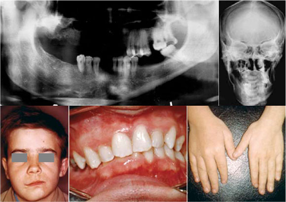
Below are images of two patients with hemifacial asymmetry causing obvious disfigurement of the face and jaws. What is your diagnosis?
INTERPRETATION
The pantomograph and posteroanterior projection (upper images) showing marked hypertrophy of the right mandible while the lower clinical pictures of another patient show hypertrophy of the soft tissues of the face and jaw and macrodactyly of the right hand. A diagnosis of hemifacial hypertrophy was made. Nearly everybody has mild facial asymmetry. In hemifacial hypertrophy, however, there is obvious noticeable disfigurement. Congenital hemifacial hypertrophy is a rare developmental disorder of unknown etiology, characterised by a marked unilateral facial asymmetry. This phenomenon was first described by Meckel1 in 1822 and first reported by Kottmeier and Wagner2 in 1839. It involves the hard (bones and teeth) and soft tissues of the face. Hypertrophy is an increase in the size of an organ or a part thereof as a result of enlargement of the component cells. Perhaps more accurately, hyperplasia is enlargement due to an increased number of cells and not their size and this may be limited to the head or may involve one half of the body. In congenital hemifacial hypertrophy, the right and left sides are almost equally involved. About two thirds of reported cases are in females. Affected patients have enlargement of one half of the head, which may present at birth or may develop later. The disproportion remains and can worsen during growth. A relationship has been reported between hemifacial hypertrophy and childhood dysplasia including Wilms tumour, hepatoblastoma, and tumours of the suprarenal gland cortex. Oral manifestations sometimes include developmental anomalies of the dentition on the affected side. Permanent teeth may show macrodontia, being up to 50% larger than those on the unaffected side. Canines, premolars, and first permanent molars appear to be the teeth most commonly enlarged. Affected teeth may develop more rapidly and erupt before those on the contralateral side. The jaws and tongue are often involved in hemifacial hypertrophy. The asymmetry usually remains constant with the end of adolescence, and as skeletal maturation occurs the condition stabilizes thereafter. Skeletal deformities have also been noted in hemifacial hypertrophy which include macrodactyly.
Reference
1. J. F. Meckel, "Ueber die seitliche Asymmetric im tierischen Korper," in Anatomische Physiologische Beobachtungen und Untersuchungen, R. Halle, Ed., p. 147, 1822. [ Links ]
2. H. L. Kottmeier and H. L. Wagner, "Über Hemihypertrophia und Hemiatrophia corporis totalis nebst spontane Extremitätengangräne bei Säuglingen im Anschluss zu einem ungewöhnlichen," Fall Acta Paediatrica, vol. 20, no. 4, pp. 530-543, 1938. [ Links ]
3. Farman AG, Nortjé CJ & Wood R E: Oral and Maxillofacial Imaging, 1st Ed, Mosby. St. Louis, Missouri 1993 p 128-129. [ Links ]
