


Services on Demand
Journal

Article
 English (pdf)
 Article in xml format
 Article references
 Send this article by e-mail
Indicators
Related links
-
 Cited by Google
 -
 Similars in Google
Share

 Permalink
SA Journal of Radiology
On-line version ISSN 2078-6778Print version ISSN 1027-202X
S. Afr. J. radiol. (Online) vol.18 n.1 Johannesburg Oct. 2014
SIGNS
Persistent hyperplastic primary vitreous - The martini glass sign
A. Fourie Bezuidenhout
Department of Radiodiagnosis, Stellenbosch University, South Africa
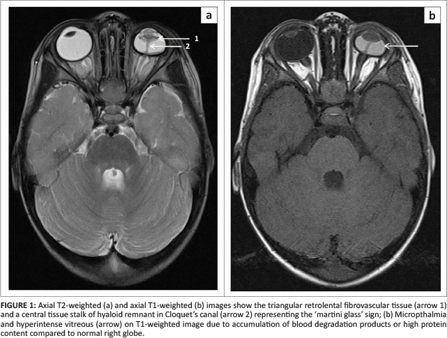
Persistent hyperplastic primary vitreous (PHPV) is a congenital lesion due to incomplete regression of the embryonic ocular blood supply (hyaloid vasculature).1 It represents 28% of childhood presentations of leukocoria and is almost always accompanied by poor vision, micropthalmia and often retinal detachment.1-2-3 The absence of ocular calcifications helps distinguish PHPV from the more common retinoblastoma.4 The appearance of PHPV has been likened to that of a martini glass. The martini glass is represented by triangular retrolental fibrovascular tissue and a central tissue stalk of hyaloid remnant extending to the optic disc in Cloquet's canal (see Figure 1a).5 The retrolental fibrovascular tissue and stalk-like hyaloid remnant are hypointense to isointense on T1- and T2-weighted images and show enhancement post contrast administration. The globe may be hyperintense on T1-weighted images; this may represent either subhyaloid or subretinal fluid with blood degradation products (methaemoglobin) or high protein content (see Figure 1b).1
Acknowledgements
Competing interests
The author declares that he has no financial or personal relationship(s) that may have inappropriately influenced him in writing this article.
References
1. Smirniotopoulos JG, Bargallo N, Mafee MF. Differential diagnosis of leukokoria: Radiologic-pathologic correlation. Radiographics. 1994;14(5):1059-1079. PMID: 7991814. http://dx.doi.org/10.1148/radiographics.14.5.7991814 [ Links ]
2. Kuker W, Ramaekers V. Persistent hyperplastic primary vitreous: MRI. Neuroradiology. 1999;41(7):520-522. PMID: 10450848. http://dx.doi.org/10.1007/s002340050796 [ Links ]
3. Sun MH, Kao LY, Kuo YH. Persistent hyperplastic primary vitreous: Magnetic resonance imaging and clinical findings. Chang Gung Med J. 2003 Apr;26(4):269-276. PMID: 12846526. [ Links ]
4. Edward DP, Mafee MF, Garcia-Valenzuela E, Weiss RA. Coats' disease and persistent hyperplastic primary vitreous. Role of MR imaging and CT. Radiol Clin North Am. 1998;36(6):1119-1131, x. PMID: 9884692. [ Links ]
5. Kaste SC, Jenkins JJ, Meyer D, Fontanesi J, Pratt CB. Persistent hyperplastic primary vitreous of the eye: Imaging findings with pathologic correlation. Am J Roentgenol. 1994;162(2):437-440. PMID: 8310942. http://dx.doi.org/10.2214/ajr.162.2.8310942 [ Links ]
 Correspondence:
Fourie Bezuidenhout
PO Box 19179
Tygerberg 7505
South Africa
Email: fouriebez@yahoo.com
Received: 07 Jan. 2014
Accepted: 16 Feb. 2014
Published: 20 June 2014
