


Services on Demand
Journal

Article
 English (pdf)
 Article in xml format
 Article references
 Send this article by e-mail
Indicators
Related links
-
 Cited by Google
 -
 Similars in Google
Share

 Permalink
South African Journal of Science
On-line version ISSN 1996-7489Print version ISSN 0038-2353
S. Afr. j. sci. vol.121 n.5-6 Pretoria May./Jun. 2025
https://doi.org/10.17159/sajs.2025/17896
RESEARCH ARTICLE
The impact of radiation dose variability on the response of radiophotoluminescence glass dosimeters - an experimental approach
Manny MathuthuI; Samuel O.O. JohnI; Dithole H. SeepamoreII; Vincent MaseleseleIII
ICentre for Applied Radiation Science and Technology (CARST), North-West University, Mmabatho, South Africa
IINational Metrology Institute of South Africa, Pretoria, South Africa
IIISteve Biko Academic Hospital, Pretoria, South Africa
ABSTRACT
The dosimetric properties of radiophotoluminescence glass dosimeters (RPLGDs) make them valuable tools for accurately measuring doses in various radiation fields. Over the years, thermoluminescence dosimeters have been used for both personal and environmental monitoring in South Africa, although they have certain limitations. We investigated the exceptional properties of RPLGDs by characterising their response to different radiation doses, using radiation sources of 60Co, 137Cs, and 241Am. The objective was to assess the ability of RPLGDs to be read multiple times without losing the original signal, and to explore their potential to replace TLDs in diverse radiation environments. A substitution method was employed to determine the reference measurements across all radiation source set-ups. In this approach, the RPLGD, serving as the unit under test, was exposed to the same dose as the ionisation chambers, which acted as reference detectors to accumulate the radiation signal, which was then corrected to determine the air kerma and absorbed dose to water. All relevant corrections affecting the unit under test response were applied to the final readings to characterise the RPLGDs, which were compared with the prescribed dose. The findings of this research are valuable to medical facilities and radiation workers as they offer both technical and economic benefits through improving the accuracy and reliability of radiation dose monitoring.
SIGNIFICANCE:
• Radiophotoluminescence glass dosimeters (RPLGDs) were successfully characterised with 60Co, 137Cs, and 241Am radiation beams.
• The glass dosimeters were annealed and irradiated at a temperature of 400 °C.
• The RPLGD calibration coefficients, air kerma rate and absorbed dose to water measurements were established.
• RPLGDs are capable of being re-read multiple times without losing any signal.
Keywords: radiophotoluminescence glass dosimeter, thermoluminescent dosimeter, air kerma, absorbed dose, radionuclide beam
Introduction
Ensuring accurate radiation dose measurement and delivery across diagnostic radiology, radiation therapy, radiation protection and environmental monitoring is a priority, especially in the fight against cancer and in safeguarding public health and the environment. Radiation dosimetry plays a pivotal role in safeguarding human health and the environment by ensuring accurate dose measurement in a wide array of applications, from cancer treatment in radiotherapy to radiation protection in industrial and environmental settings.1-3 Specialised dosimeters, such as radiophotoluminescence glass dosimeters (RPLGDs), are indispensable tools for quantifying and monitoring radiation doses across these various fields. Dosimetric quantities, such as air kerma and absorbed dose, are used to determine the amount of energy imparted by a radiation beam. Air kerma is a measure of the kinetic energy released to matter.
The determination of physical quantities like air kerma and absorbed dose to water requires both the radiation source and measuring instruments. In this study, RPLGDs were employed for calibration purposes. The radiation sources used, as recommended by the International Organization for Standardization (ISO 4037-1), were 60Co, 137Cs and 241Am, with their respective energies of 1252 keV (the mean of 1173 keV and 1332 keV), 662 keV and 59.5 keV.4,5
Ionising radiation can be an invaluable tool for various medical and industrial applications, but it also poses significant risks when it interacts with matter. While harmful to living organisms, radiation has proven beneficial in treatments such as cancer therapy.2,6 To detect different types of radiation, different radiation instruments are used. Different types of radiation - gamma rays, X-rays, beta particles and alpha particles - can deposit energy in matter.7,8 In this study, the focus was on the interaction of gamma radiation with luminescent materials. The material under investigation in this study was the RPLGD, a member of the family of passive dosimeters, which also includes thermoluminescence dosimeters (TLD) and optical stimulated luminescence dosimeters (OSLD).9 We explored the response of RPLGDs to gamma radiation, with an emphasis on the ability of the RPLGDs to measure air kerma and absorbed dose to water. For more than 50 years, scientists have worked on developing RPLGDs. They were first introduced in 1947 with a suboptimal luminescent material and readout technique. However, in 1950, a new RPLGD, using a glass compound as the luminescent material (with a pulsed ultraviolet laser beam to excite the glass compound, thereby causing an orange luminescence to be emitted), was developed to address the limitations of TLDs.10,11
The excitation source was switched from ultraviolet (UV) light to pulsed UV laser beams. When a silver-activated phosphate glass compound of the RPLGD is exposed to ionising radiation, stable colour centres form. These colour centres increase in number as radiation intensity rises. Upon irradiation, the colour centres are excited by a pulsed UV laser beam, and emit orange light with wavelengths between 600 nm and 700 nm - a phenomenon known as radiophotoluminescence.12 The radiation dose received by glass dosimeters is directly proportional to the intensity of the orange light emitted by the RPLGD. One key advantage of the RPLGDs is that their signal can be read repeatedly without losing the original information, unlike TLDs, which can only be read once.
TLDs are the most widely used traditional passive dosimeters in radiation protection for personal and environmental monitoring. However, TLDs and optical stimulated luminescence dosimeters employ different excitation methods and readout techniques. The luminescent materials that constitute the solid structure of TLDs are typically phosphors, such as calcium fluoride (CaF) or lithium fluoride (LiF). When radiation interacts with the solid crystal, it deposits energy to the phosphor, ionising the material.13 The energy deposited by radiation is retrieved by annealing the dosimeter, exciting the electrons to a higher energy state. The excited electrons then return to the ground state, releasing energy that corresponds to the absorbed dose. However, a major limitation of TLDs is their inability to retain and re-read a signal once it has been read, as the trapped radiation energy dissipates after a single readout. This challenge poses a significant issue in radiation protection and its applications, particularly when repeated dose measurements are required, with high precision. In contrast, RPLGDs offer distinct advantages, including excellent reproducibility of the readout values, long-term stability with fading effects of less than 1% over 30 days, and low energy dependence for photons in the range of 0.03-1.3 MeV. Additional advantages include a good dose linearity (0.00001-0.5 Gy) and the ability to repeatedly read out the dosimeter's signal, making RPLGDs a promising alternative to TLDs.14,15
In this study, we aimed to address the challenges associated with TLDs by exploring the potential of RPLGDs as an effective alternative passive dosimeter. The primary objective was to develop reference measurements for air kerma and absorbed dose to water for RPLGDs using three radiation sources: 60Co, 137Cs and 241Am. These measurements will support the development of a viable alternative to the limitations posed by TLDs, which are predominantly used in medical facilities and by radiation workers in South Africa, as recommended by the Department of Health (DoH) and the South African Health Products Regulatory Authority. By providing baseline reference data, this study will facilitate the adoption of RPLGDs in medical facilities and among radiation workers in South Africa, addressing the critical issue of signal loss in accumulated radiation dose measurements. The findings of this research are valuable to medical facilities and radiation workers, as they offer both technical and economic benefits through improving the accuracy and reliability of radiation dose monitoring.
Experimental methods
The materials and methods used in this study were based on the procedures developed in the Dosimetry Standards Laboratory of the National Metrology Institute of South Africa in Pretoria. Two reference radiation detectors were employed to obtain reference measurements for characterising RPLGDs. These were the Physikalisch-Technische Werkstatten GmbH (PTW) spherical ionisation chamber (1000 cc) and PTW cylindrical ionisation farmer chamber (0.6 cc). The RPLGD model used was the RPL glass element of the International Atomic Energy Agency (type FD-R1.5(12)-7). A total of 58 RPLGDs were used in this study, each with a diameter of 1.5 mm and a length of 12 mm.
These ionisation chambers served as reference detectors to determine the air kerma rate from three radiation sources: 60Co, 137Cs and 241Am, all of which are located inside irradiators. The RPLGDs were characterised against the reference detectors using the substitution method, whereby ionisation was exchanged with RPLGDs under identical set-up conditions. The Dose Ace reader, capable of reading doses from 0.00001 Gy to 10 Gy (with an extended range of up to 100 Gy), was used to read the glass dosimeters. This reader employs a pulsed UV laser to excite electrons in the RPLGDs. When the silver phosphate glass is excited by the pulsed UV laser, it emits visible orange light (600-700 nm) and returns to its original colour centre. To extract meaningful information from the radiation counts (which are linearly proportional to absorbed radiation), the calibration coefficient for each radiation source per glass dosimeter and relevant correction factors were applied to the recorded radiation counts.11
Experimental set-up
The experimental set-up involved positioning the radiation sources at specific distances from the dosimeters placed in their respective phantoms, based on their activity. The distances were set at 100 cm for the 60Co source, 200 cm for the 137Cs source and 50 cm for the 241Am source, as shown in Figure 1a-f. Two laser beams, mounted on the wall and aligned perpendicular to each other, were used to position the farmer ionisation chamber and the RPLGD.
The calibration coefficient (ND, W) for the 60Co source was obtained from the calibration certificate (No.65 (2023) ion chamber in 60Co, PTW30010-132), which was calibrated in water at the Primary Standards Laboratory at the BIPM (International Bureau of Weights and Measures) in France. The calibration coefficient, (ND, W), was given as 5.32E+07 Gy/C. The farmer chamber has a volume of 0.6 cm3 (or 1 L) and an inner diameter of 6.1 mm. The calibration coefficient (NK) for the 137Cs source was 2.518E+04 Gy/C, as stated in its calibration certificate. The chamber has a volume of 1000 cm3 and a diameter of 140 mm. The calibration coefficient (NK) for the 241Am source was 2.513E+04 Gy/C. The equipment used was verified and calibrated to ensure traceability of air kerma measurements to national and international standards. Additionally, the absorbed dose to water could be traced back to the BIPM.
Before irradiation, the glass dosimeters were annealed at 400 °C for 2 h, then allowed to cool for at least 2 h. This process allowed the electrons to return to the valence band. The background counts recorded after annealing were subtracted from the absorbed radiation counts. After irradiation, the glass dosimeters were pre-heated at 70 °C for at least 1 h in an oven to excite the captured electrons into the conduction band. They were then cleaned in an ultrasonic bath with ethanol to remove any contamination.
A read-out magazine (numbered 018) was used to process the dosimeters. The magazine has 20 positions in which the RPLGDs were inserted for reading. The 20 RPLGDs were irradiated with 2 Gy from the 60Co beam and read in all 20 positions. The global average of all the readings was calculated, and each reading was normalised to the global average.
Prescribed radiation doses used to irradiate RPLGDs
Prescribed doses refer to the absorbed dose expected to be read on the RPLGDs after their irradiation with respective radiation sources. Each source has a reference dose, which was used to calculate the calibration coefficient of the glass dosimeter. The reference doses are highlighted in bold in Table 1 for each source.

The calibration coefficient for the glass dosimeter in the 60Co beam was determined for the absorbed dose to water using solid water measurements, with the glass dosimeter placed inside the solid water phantom. The radiation counts obtained when the glass dosimeters were irradiated at a distance of 100 cm, with a prescribed radiation dose of 2.0 Gy, were taken as the reference radiation dose (M ref 2.0 Gy). The calibration coefficient NRPLGD(60Co) was calculated using Equation 1:
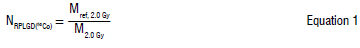
where M2.0Gy is the average of all radiation counts from six glass dosimeters exposed to the reference dose of 2.0 Gy.
The air kerma calibration coefficient (NRPLGD(137Cs)) for the glass dosimeter with the Perspex rod was calculated using Equation 2. The radiation count obtained when the glass dosimeter was irradiated at 200 cm with a prescribed radiation dose of 0.020 Gy was taken as the reference radiation dose (Mref, 0.02 Gy).
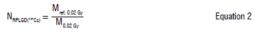
where M0.02 Gy is the average of all radiation counts from five glass dosimeters exposed to a reference dose of 0.02 Gy.
Similarly, the air kerma calibration coefficient NRPLGD(241Am) for the glass dosimeter with the Perspex rod was calculated using Equation 3. The radiation count obtained when the glass dosimeters were irradiated at 50 cm with a prescribed radiation dose of 0.00002 Gy was taken as the reference radiation dose Mref, 0.00002 Gy
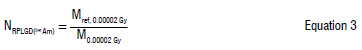
where M0.00002 Gy is the average of all radiation counts from three glass dosimeters exposed to the reference dose irradiated with a prescribed dose of 0.00002 Gy (i.e. the average radiation counts of the three glass dosimeters exposed to the reference dose)
These calibration coefficients were then used to determine the air kerma rate by Equation 4.16
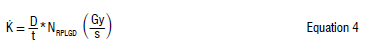
where D is the absorbed dose read from the RPLGD, t is the irradiation time, and NRPLGD is the dosimeter's calibration coefficient forthe radiation source.
Temperature, pressure and charge measurements
The detectors used in this study were corrected for temperature and pressure, with humidity monitored under allowable working conditions in the laboratory.17 Temperature and pressure measurements were recorded before irradiation (T1 and P1) and after irradiation (T2 and P2). Equations 5 and 6 were used to obtain interpolated temperature and pressure values, found between two instrument readings indicated on the calibration certificate.
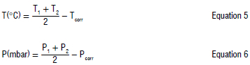
where Tcorr and Pcorr are the temperature and pressure corrections between the reference measurements and the instrument's readings obtained during calibration.
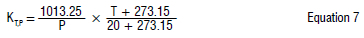
Equation 7 was used to calculate the temperature and pressure correction factors, with results presented in Table 2.
Charge background measurements were recorded to quantify chamber leakages when the radiation source was off (i.e. pre- and post-irradiation measurements), as well as during irradiation. Background measurements were recorded before and after irradiation correction for any leakage in the ionisation chambers. The averages of the pre-irradiation (Qpre), post-irradiation (Qpost) and irradiation measurements (Qirr) were calculated using Equation 8. The leakage, Qleakage, was obtained from the average of Qpre and Qpost.
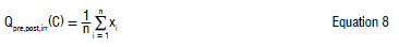
where n is the total number of charge measurements and xi is the individual charge measurement.
The irradiation time T, the ratio of absorbed dose to dose rate, was recorded in seconds, except for the 60Co source, where time was recorded in minutes as entered into the irradiation software. The intrinsic effect was tested using the ionisation chambers, with no signal detected during intrinsic time measurements.
Results and discussion
Figure 2 shows the magazine corrections plotted against the 20 magazine slots, illustrating the relationship between the corrections applied to the RPLGDs and their respective positions in the magazine. The plot features three distinct curves: the actual data curve (blue), the curve fit (green) and the Gaussian curve fit (red). The actual data curve represents the raw, experimental measurements taken from the dosimeters positioned in various slots of the magazine. The curve fit was applied to the actual data in order to approximate the relationship between the magazine corrections and slot numbers. The curve is based on an algorithm that minimises the difference between the fitted curve and actual data. The Gaussian curve fit (or bell-shaped) was applied and reflects the nature of variation in the magazine corrections across the magazine slots. A second-order polynomial was also fitted to determine the function used in the calculation of magazine correction factors (red curve). The quadratic term (-0.0004x2) indicates that the correction values follow a parabolic pattern. The linear term (0.0087x) slightly shifts the curve, influencing the slope of the correction values, while the constant term (0.9658) represents the baseline correction factor for the central slot (x = 0). The use of the Gaussian curve fit helps to validate that the magazine corrections exhibit a symmetrical, bell-shaped distribution around the central position. This suggests that the RPLGDs' performance is generally more consistent in the centre slots and deviates towards the edges. This model provides a reliable understanding of how the magazine slot number affects the correction values applied to the dosimeter readings, which is crucial when standardising and calibrating dosimeters across varying slot positions in the magazines. The magazine correction factors are used throughout the measurements for position correction of the magazine reader. These factors played a significant role in optimising the results.
Table 2 shows the temperature and pressure measurements for the 60Co, 137Cs and 241Am sources. The temperature range for the radiation sources is from 18.377 °C to 20.171 °C, while the pressure range spans from 868.40 mbar to 874.46 mbar. The correction factors for temperature and pressure range from 1.1523 to 1.1658. The highest value of temperature was recorded for the 137Cs source, while the highest pressure was recorded for the 60Co source. The relative humidity recorded for the sources was 54.20%RH, 42.40%RH and 50.00%RH, for 60Co, 137Cs and 241Am, respectively. Although the measurements were not corrected for humidity, the temperature and pressure measurements were corrected.
The RPLGDs responded well to the environmental conditions of temperature and pressure. The annealing temperature of 400 °C and subsequent irradiation were sufficient for appropriate characterisation of the RPLGDs. The importance of temperature and pressure measurements is central to the sensitivity of the glass dosimeter, which aligns with the findings of Sato et al.15
Figure 3 shows the charge measurements recorded pre- and post-irradiation for the radiation sources for the ionisation chamber and RPLGDs. The average charge (Q irr) collected from the sources ranged from -1.31750E-10 C for 241Am to -7.24740E-9 C for the 60Co source, indicating a reasonable charge range for detection by the RPLGD. The wide range of charges observed reflects the varying intensity of the radiation fields generated by each source, as well as the sensitivity of the RPLGDs to these radiation levels.
The corrected charge measurement values for the sources, with respect to the RPLGDs, ranged from -1.532E-10 C for 241Am to -1.047E-09 C for 137Cs. These corrected values provide a more accurate reflection of the dosimeter's response to radiation, considering potential influences such as environmental factors, detector calibration, and other adjustments.
It is worth noting that the negative charge values are consistent with the expected behaviour of ionisation chambers and RPLGDs, which generate a negative charge when exposed to ionising radiation. The relatively higher charge for the 60Co source compared to 241Am can be attributed to the higher energy and intensity of the 60Co radiation, which resulted in greater ionisation and, therefore, a higher recorded charge.
The charge measurements are critical for determining the dose rates for each radiation source. The relationship between the charge collected and the radiation dose rate is well established, and these charge measurements provide essential data for calculating the absorbed dose delivered by the radiation sources. The results are further used to determine the dose rate for the various radiation sources in relation to the RPLGDs, which is key for evaluating the dosimeter's accuracy and effectiveness.3 The subsequent calculations and analysis will help assess the feasibility of using RPLGDs as a reliable alternative to traditional dosimetry methods, particularly in clinical settings.
Additionally, comparing the charge measurements from the ionisation chamber and the RPLGDs allows for an evaluation of the consistency and reliability of the RPLGDs. If the charge measurements between the two methods are consistent, it would support the case for using RPLGDs in routine dosimetry applications. If discrepancies are observed, further investigation into the sources of error, such as calibration or environmental factors, would be necessary.14
The charge measurements serve as a critical component in validating the performance of the RPLGDs in relation to traditional ionisation chambers, providing valuable insight into suitability for practical dosimetry applications.10
The charges measured were used to obtain the dose rate for the various sources and in relation to the RPLGD.
Figure 4 shows the irradiation times and corresponding prescribed doses for various sources on the RPLGD, demonstrating the optimised response of the glass dosimeter. The results indicate that 60Co yielded higher irradiation times (in seconds), corresponding to its higher radiation levels, while 241Am presented shorter irradiation times, also measured in seconds. The compatibility of the measurements with the time, along with the re-read results, confirms that the RPLGD is sufficiently sensitive to be used in place of TLDs, thereby addressing their limitations.
The dose rate and corrected charge (Q) for the respective sources on the RPLGD are 0.538 Gy/s, -8.38463E-09 C for 60Co, 8.785E-08 Gy/s, -1.04666E-09 C for 137Cs, and 6.416E-07 Gy/s, -1.53186E-10 C for 241Am. The values are presented in a simplified format for ease of calculating the time set on the irradiator during RPLGD irradiation. As expected, 60Co had the highest dose rate, while 241Am had the lowest. The results align with the prescribed doses and source strengths of the radiation sources used. Furthermore, the corrected charge values show negligible error percentages, indicating improved accuracy in the charge measurements.
The calibration coefficients of the RPLGDs were determined using Equations 1-3, with the radiation counts taken at the reference doses of 2.0 Gy (for 60Co), 0.02 Gy (for 137Cs) and 0.00002 Gy (for 241Am). The mean values of the calibration coefficients, along with their respective uncertainties, were found to be 9.347E-07 ± 4.030E-09 Gy/C for 60Co, 7.962E-07 ± 6.048E-10 Gy/C for 137Cs and 2.056E-08 ± 5.323E-11 Gy/C for 241Am. It is noteworthy that the calibration coefficients for the farmer ionisation chamber, which was calibrated in water for the radiation sources, were found to be higher than those of the RPLGD. This suggests that the RPLGD has a unique property that enables it to measure very low radiation doses, as low as 0.00001 Gy, owing to its higher sensitivity compared to the ionisation chamber.
Table 3 summarises the absorbed dose and the air kerma rate measured for the RPLGDs under irradiation from the three radiation sources: 60Co, 137Cs, and 241Am. The table shows the results for each RPLGD, along with the corresponding uncertainties at k = 2, based on measurements of absorbed dose (Gy) and air kerma rate (Gy/s).
From Table 3, the absorbed dose values for 60Co, 137Cs, and 241Am radiation sources show a clear trend in increasing values with respect to the radiation energy and exposure levels. For 60Co, the absorbed doses range from 0.1990 ± 0.005 Gy to 9.1475 ± 0.210 Gy with air kerma rates of 1.41E-08 Gy/s. Similarly, for 137Cs, the absorbed doses range from 0.0099 ± 2.49E-04 Gy to 0.0415 ± 1.67E-04 Gy, with corresponding air kerma rates ranging from 6.94E-12 Gy/s to 7.25E-12 Gy/s. For 241Am, the absorbed doses ranged from 1.91E-05 ± 1.15E-07 Gy to 13.46E-05 ± 6.66E-07 Gy, with air kerma rates ranging from 4.54E-16 Gy/s to 4.57E-16 Gy/s.
The data show a high level of consistency between the absorbed doses measured by the RPLGD and the prescribed doses for the three radiation sources. The uncertainties at k = 2 were minimal, confirming the excellent precision of the RPLGD. The R2 value of 0.971 indicates a strong correlation between absorbed doses and air kerma rates, suggesting a good overall response from the RPLGD. The air kerma rates calculated for the different radiation beams, as presented in Table 3 (columns 3, 5 and 7), show similar values for each respective radiation source. The average air kerma rates for each source are as follows: 1.40E-08 ± 1.60E-10 Gy/s for the 60Co radiation beam, that of 137Cs is 7.04E-12 ± 1.10E-13 Gy/s and that of 241Am is 4.75E-16 ± 1.76E-17 Gy/s. These values reflect the different radiation energies associated with each source, as well as the prescribed doses. The RPLGD consistently provided accurate and sensitive measurements, even at very low dose levels, demonstrating its potential for use in radiation monitoring and dosimetry applications. Therefore, it can be concluded that the RPLGD demonstrated excellent performance in measuring absorbed doses and air kerma rates for 60Co, 137Cs and 241Am radiation sources. Its ability to measure low doses accurately, its high sensitivity, and repeatable readings make it a strong alternative to traditional dosimetry methods, such as ionisation chambers and TLDs. The data obtained from RPLGDs align well with the prescribed radiation doses, further supporting their use in precise radiation monitoring applications.
Conclusion
The characterisation of RPLGDs was aimed at quantifying their response when exposed to gamma radiation from 60Co, 137Cs and 241Am sources. This study stemmed from the use of TLDs, which are widely employed by radiation workers in South Africa. The use of TLDs is mandated by the South African Health Products Regulatory Authority (SAHPRA) to ensure proper radiation monitoring of workers handling radiation sources in their activities.
When the RPLGDs were exposed to various radiation doses from different radiation beams, with correction factors applied to all parameters with significant influence, the final measurement results were found to be in close agreement with the prescribed doses. The successful characterisation of RPLGD using 60Co, 137Cs and 241Am radiation beams has been demonstrated, with calibration coefficients, air kerma rates, and absorbed dose to water measurements all being established.
The traceability of the reference measurements was ensured by utilising the calibration coefficients of ionisation chambers, which provided the foundation for the reference measurements used in the characterisation of the glass dosimeters for air kerma and absorbed dose to water. Reference detectors were employed to determine the dose rates at the prescribed reference distances, which in turn facilitated the calculation of the necessary irradiation times for the RPLGDs. This process highlighted the unique properties of the RPLGDs studied, especially in terms of their sensitivity and reliability.
Based on the results, the RPLGDs demonstrated excellent response characteristics and were capable of being re-read multiple times without any noticeable signal loss, showcasing their robustness and accuracy. This suggests that RPLGDs hold great promise for long-term radiation monitoring applications.
Further studies will focus on exploring the energy dependence of the RPLGDs, investigating the fading effects, and evaluating the long-term stability and depletion of the dosimeters. These investigations will further contribute to enhancing the dosimetric performance and expanding the practical applications of RPLGDs in radiation monitoring.
Acknowledgements
We acknowledge the National Metrology Institute of South Africa (NMISA) for providing access to equipment and facilities for the experiment and the International Atomic Energy Agency (IAEA) for providing the radiophotoluminescence glass dosimeters.
Data availability
The data supporting the results of this study are available upon request to the corresponding author.
Declarations
We have no competing interests to declare. ChatGPT was used to correct spelling and grammatical errors.
Authors' contributions
M.M.: Conception and design of the study, data collection, data analysis, supervision. S.O.O.J.: Conception and design of the study, data analysis, writing - the initial draft. D.H.S.: Conception and design of the study, data collection, data analysis, writing - the initial draft. V.M.: Conception and design of the study, data collection, data analysis, supervision. All authors read and approved the final manuscript.
References
1. International Atomic Energy Agency (IAEA). Radiation protection and safety of radiation sources: International basic safety standards. No GSR Part 3. Vienna: IAEA; 2014. https://doi.org/10.61092/iaea.u2pu-60vm [ Links ]
2. Vaz P. Radiation protection and dosimetry issues in the medical applications of ionizing radiation. Radiat Phys Chem. 2014;104(1):23-30. https://doi.org/10.1016/j.radphyschem.2014.02.007 [ Links ]
3. Adliené D, Adlyté R. Dosimetry principles, dose measurements, and radiation protection. In: Sun Chmielewski AG, editors. Applications of ionizing radiation in materials processing. Warsaw: Institute of Nuclear Chemistry and Technology; 2017. p. 55-80. [ Links ]
4. Dietze G. Dosimetric concepts and calibration of instruments: International Radiation Protection Association (IRPA) [document on the Internet]. c2001 [cited 2023 Jun 18]. Available from: https://www.irpa.net/irpa10/pdf/E03.pdf [ Links ]
5. International Organization for Standardization (ISO). X and gamma reference radiation for calibrating dosemeters and dose rate meters and for determining their response as a function of photon energy: Radiation characteristics and production methods [document on the Internet]. c1996 [cited 2025 Jan 31]. Available from: https://www.iso.org/obp/ui/#iso:std:iso:4037:-1:ed-1:v1:en [ Links ]
6. Hooshang N, Shuzo U, Dimitris E. Interaction of radiation with matter. New York: CRC Press; 2012. https://doi.org/10.1201/b12109 [ Links ]
7. John SOO, Usman IT, Akpa TC, Abubakar SA, Ekong GB. Natural radionuclides in rock and radiation exposure index from uranium mine sites in parts of Northern Nigeria. Radiat Prot Environ. 2020;43(1):36-43. https://doi.org/10.4103/rpe.RPE_7_20 [ Links ]
8. Mathuthu M, Uushona V, Indongo V. Radiological safety of groundwater around a uranium mine in Namibia. Phys Chem Earth. 2021;122(1), Art. #102915. https://doi.org/10.1016/j.pce.2020.102915 [ Links ]
9. Akselrod M, Bøtter-Jensen L, McKeever S. Optically stimulated luminescence and its use in medical dosimetry. Radiat Meas. 2006;41(1):S78-S99. https://doi.org/10.1016/j.radmeas.2007.01.004 [ Links ]
10. Rah J-E, Hong J-Y, Kim G-Y, Kim Y-L, Shin D-O, Suh T-S. A comparison of the dosimetric characteristics of a glass rod dosimeter and a thermoluminescent dosimeter for mailed dosimeter. Radiat Meas. 2009;44(1):18-22. https://doi.org/10.1016/jradmeas.2008.10.010 [ Links ]
11. Seepamore DH. Quantification of radio-photoluminescence glass dosimeter with different radionuclide beams [MSc dissertation]. Mmabatho: North-West University; 2021. Available from: https://repository.nwu.ac.za/bitstream/handle/10394/38202/18029701%20seepamore%20dh.pdf?sequence=1 [ Links ]
12. Miyamoto Takei Nanto H, Kurobori T, Konnai A, Yanagida T, et al. Radiophotoluminescence from silver-doped phosphate glass. Radiat Meas. 2011;46(12):1480-1483. https://doi.org/10.1016/j.radmeas.2011.05.048 [ Links ]
13. Weihai Z, Weiqi L, Guoying Z, Gang H, Guocai M. Comparisons of dosimetric properties between radiophotoluminescent glass rod detector and thermoluminescent detectors [document on the Internet]. c2007 [cited 2025 Jan 31]. Available from: https://www.osti.gov/etdeweb/biblio/21118320 [ Links ]
14. Huang DY, Hsu S-M. Radio-photoluminescence glass dosimeter (RPLGD. In: Gali-Muhtasib H, editor. Advances in cancer therapy. London: InTechOpen; 2011. p. 553-568. https://doi.org/10.5772/23710 [ Links ]
15. Sato F, Zushi N, Nagai T, Tanaka T, Kato T, Yamamoto T, et al. Development of radiophotoluminescence glass dosimeter usable in high temperature environment. Radiat Meas. 2013;53-54(1):8-11. https://doi.org/10.1016/j.radmeas.2013.03.016 [ Links ]
16. International Atomic Energy Agency (IAEA). Dosimetry in diagnostic radiology: An international code of practice. TRS No. 457 [document on the Internet]. c2007 [cited 2025 Jan 31]. Available from: https://www.iaea.org/publications/7638/ [ Links ]
17. South African National Accreditation System (SANAS). TR 18-02 [document on the Internet]. c2014 [cited 2025 Jan 31]. Available from: https://www.sanas.co.za/Publications%20and%20Manuals%20Files/TR%2018-02.pdf [ Links ]
 Correspondence:
Samuel John
Email: samjoh2014@gmail.com
Received: 29 Apr. 2024
Revised: 05 Feb. 2025
Accepted: 19 Feb. 2025
Published: 29 May 2025
Editors: Michael Inggs,Thywill Dzogbewu
Funding: None
