


Serviços Personalizados
Journal

Artigo
 Inglês (pdf)
 Artigo em XML
 Referências do artigo
 Enviar este artigo por email
Indicadores
Links relacionados
-
 Citado por Google
 -
 Similares em Google
Compartilhar

 Permalink
SA Journal of Radiology
versão On-line ISSN 2078-6778versão impressa ISSN 1027-202X
S. Afr. J. radiol. (Online) vol.19 no.1 Johannesburg 2015
https://doi.org/10.4102/SAJR.v19i1.834
QUIZ CASE
E.B. ArkinkI, II; J.H.M. FrijnsIII; B.M. VerbistI, IV
IDepartment of Radiology, Leiden University Medical Center, The Netherlands
IIDepartment of Radiology, Medical Center Haaglanden, The Netherlands
IIIDepartment of Otorhinolaryngology, Leiden University Medical Center, The Netherlands
IVDepartment of Radiology, Radboud University Nijmegen Medical Center, The Netherlands
A 46-year-old man presented to the Department of Otolaryngology with congenital deafness of unknown cause. With the help of hearing aids (which did not provide any speech understanding in the sound-only condition) and by mastering speech reading, he had learned to speak quite fluently at a school for deaf and hearing-impaired children. Despite these capabilities, he came to seek advice about cochlear implants, as he would like to communicate more conveniently with his family, of whom none had significant hearing loss.
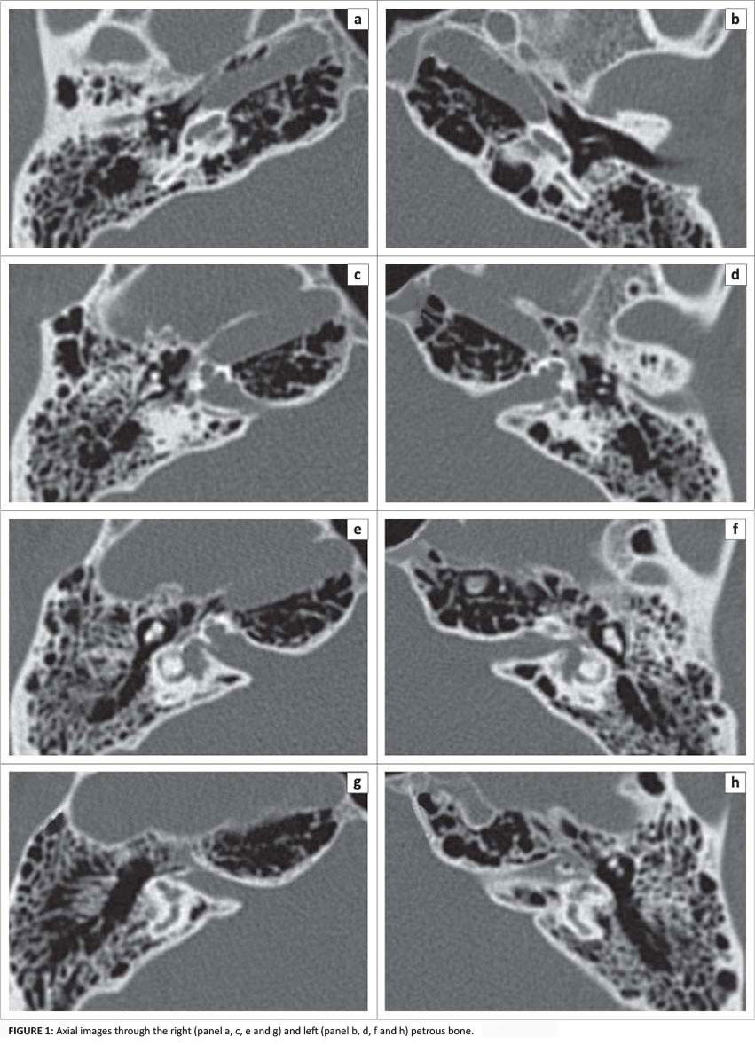
On physical examination, he was completely deaf and had adapted to major vestibular deficits (i.e. bilateral vestibular areflexia). The following CT scan of the petrous bone (Figure 1) and MRI images of the cerebellopontine angle (Figure 2) were obtained.
 Correspondence:
E.B. Arkink
Albinusdreef 2
2333 ZA Leiden, Netherlands
Email: e.b.arkink@lumc.nl
